Plasmodium falciparum - Life Cycle
Life Cycle of Plasmodium falciparum
The life cycle of Plasmodium falciparum shows an alternation of generation accompanied by an alternation of hosts. In the human host, the asexual cycle (schizogony) takes place inside the erythrocytes while the sexual cycle (sporogony) takes place in the female Anopheline mosquito.
Human Cycle
A bite by a female Anopheline mosquito during the blood meal injects the Plasmodium falciparum sporozoites into the capillaries of the skin
these parasitic sporozoites enter the blood circulation and within less than 30 minutes, they enter the host liver tissue cells
There are two main stages of development in the human host:
Exo-erythrocytic schizogony (in the liver)
Erythrocytic schizogony and gametocytogenesis (in RBC)
Exo-erythrocytic schizogony
the Plasmodium falciparum sporozoites are covered with circumsporozoite protein which facilitates the invasion of host liver cells and not other host tissues
the circumsporozoite protein binds specifically and non-covalently with receptors present hepatocyte cell membrane’s basolateral area
these sporozoites undergo asexual reproduction called pre-erythrocytic or primary exo-erythrocytic (EE) schizogony which transforms sporozoites into trophozoites
these trophozoites mature after a few days by feeding on host cell cytoplasm and the disappearance of organelles of the apical complex
multinucleate liver-stage schizonts (EE schizonts) are developed with the formation of numerous daughter nuclei in trophozoites
mature schizonts are multinucleated, spherical, and measure more than 60μm and each contains 2,000 to 50,000 uninucleate merozoites
Finally, mature EE schizonts and enlarged liver cells rupture, releasing thousands of merozoites into the host bloodstream
only a single cycle of primary exo-erythrocytic schizogony takes place in Plasmodium falciparum and is completed in 6 days
unlike in P. vivax and P. ovale, there is no secondary exo-erythrocytic schizogony and no formation of hypnozoites and there is no true relapse
Plasmodium falciparum merozoites after entering the RBC never invade the liver and thus exo-erythrocytic forms are always absent in the blood induced malaria such as during blood transfusion
Although there is no true relapse of Plasmodium falciparum infection, recrudescence of the disease may occur 1-3 years after remission of the disease
the recrudescence occurs due to the persistence of a small number of merozoites in the infected host RBC
Erythrocytic schizogony
the merozoites released from liver cells into the blood attach and invades the RBC
any age of RBC, as well as reticulocytes, are infected by Plasmodium falciparum merozoites
the invasion occurs via the apical ends of trophozoites when the major surface glycoprotein and other sialoproteins attach to the RBC membrane
in the next step, the merozoites lie within an intra-erythrocytic parasitophorous vacuole
the host hemoglobin and RBC enzymes influence the maturation of merozoites infecting the RBC but this step is suppressed if fetal hemoglobin and few other hemoglobin are present
the merozoites transform into young trophozoites or ring forms and feed on hemoglobin by consuming RBC cytoplasm
as an end, a metabolic product, a malaria pigment, and a compound of haematin and ferric acid called hemozoin are produced
the trophozoites undergo mitosis followed by division of the cytoplasm to form mature schizonts
each mature schizont contains 8-32 merozoites as well as hemozoin which eventually rupture merozoites into the blood circulation
these schizonts infect new RBCs within seconds and the cycle of schizogony repeats
in Plasmodium falciparum erythrocytic schizogony takes place inside capillaries and vascular beds of internal organs (not peripheral blood) and completes in 48 hours
Gametocytogenesis
after a few cycles of merozoites to erythrocytic schizonts to merozoites, some of the merozoites develop into gametocytes- the macrogametocytes and the microgametocytes – in the RBC of the bone marrow and spleen
gametocytogenesis process is completed in 96 hours
gametocytes in early forms are irregular in shape which eventually turns into crescent-shape
hemozoin granules in Plasmodium falciparum gametocytes are present in the central part of the cytoplasm- surrounding the nucleus
mature gametocytes are present in peripheral blood
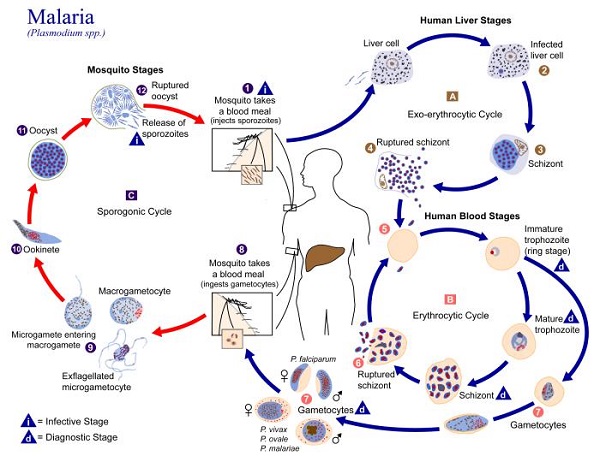
Figure: Plasmodium falciparum - lifecycle (Source: CDC)
Mosquito cycle
The sporogony or sexual life cycle of Plasmodium falciparum takes place in the female Anopheline mosquitoes and is completed in 9 days to 10 days.
Sporogony
both the macrogametocytes and microgametocytes are ingested by the female Anopheline mosquito during a blood meal from infected humans
after reaching the mid-gut (stomach) of the mosquito, the male gametocyte (microgametocyte) undergoes exflagellation, which is the transformation by rapid division
* exflagellation involves microgametocytes becoming extra cellular followed by repeated division of the nucleus within 10-12 minutes to form 6-8 daughter nuclei
* eventually, each nuclei becomes surrounded by a developing axoneme
* Finally, the microgametocyte rupture to release 6-8 daughter nuclei- with each nuclei containing an axoneme bud which later becomes microgametes
these Plasmodium falciparum microgametes are highly motile sperm-like organisms with a single flagella
the female gametocyte (macrogametocyte) matures by a single process of nuclear reduction and extension of polar bodies
each macrogametocyte can only give rise to only one macrogamete
the male gametocyte nucleus fuses with the female gametocyte nucleus through the process of fertilization and a zygote is produced
the zygote is formed within 20 to 120 minutes from the blood meal by the vector mosquito
the zygote then lengthens and extends to form a motile ookinete which is more slender in P. vivax than in other Plasmodium spp
these ookinetes penetrate the gut wall of the mosquito host, secrets a thin wall, and grow into a sphere (6μm to 12 μm in diameter) which is termed an oocyst
the oocyst contains a number of sporoblasts (haploid nucleated masses)
these sporoblasts then divide repeatedly to form sporozoites which are in turn released into the hematocele of the mosquito after rupturing of oocyst – the process called sporogony
the Plasmodium falciparum sporozoites, which are infectious to humans, migrate to the salivary glands
sporozoites are injected into the human host when the mosquito takes a blood meal from a healthy human and the life cycle of Plasmodium falciparum is continued