Schistosoma haematobium - Laboratory Diagnosis, Treatment, Prevention, Control
Laboratory diagnosis of Schistosoma haematobium
Diagnosis of Schistosoma haematobium includes cystoscopic examination and digital examination of the rectum- especially in patients of endemic areas presenting macroscopic haematuria.
In a laboratory, diagnosis of schistosomiasis is done by demonstration of typical non-operculate terminal-spined eggs.
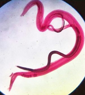
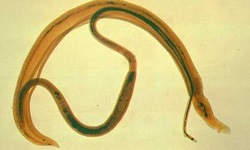
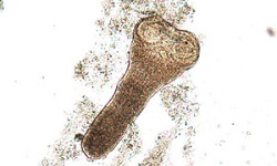
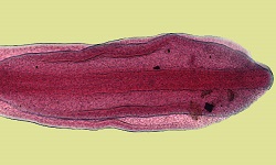
Image: adult Schistosoma haematobium - male and female (Source: Springer Link)
Samples
Urine (most common)
* Since the proportion of blood fluke eggs is excreted the most between 10 am and 2 pm, urine is collected at that time.
blood (serum)
feces (if the intestine is involved)
rectal biopsy (if the intestine is involved)
aspirations (from proctoscopy, cystoscopy)
Microscopy
Microscopy of urine samples is the most common method. Following concentration by centrifugation or membrane filtration, the residue is microscopically examined.
If urine collected within 24 hours gives the result of more than 50 eggs per 10ml of urine, infection is determined as severe while those of less than 50 eggs per 10ml of urine is considered as light infection.
Urine microscopy is also done to check for the effectiveness of treatment through egg viability tests. To test the effectiveness, urine is mixed with distilled water and observed for hatching miracidia. If eggs are viable, miracidia hatch rendering the treatment ineffective.
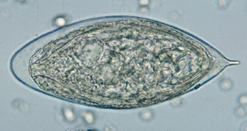
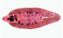
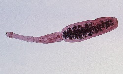
Image: S. haematobium egg (Source: CDC)
Membrane filtration
urine (10ml) is passed through a nucleophile membrane filter (13mm diameter with 8μm pre)
part of the membrane holding residue is microscopically examined at 40x magnification
presence of terminal spined eggs confirms infection by Schistosoma haematobium
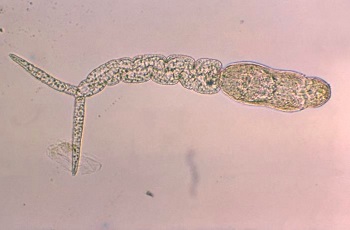
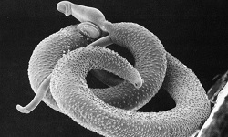
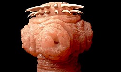
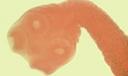
Image: S. haematobium cercaria (Source: Lecturio)
Detection of antibodies
Serodiagnosis methods can be used to demonstrate specific circulating antibodies in the patient's serum. It uses purified schistosome antigens to antigen fractions with specific antigenic determinants.
These tests are mostly used to detect infection during the pre-patent period, chronic infection, and ectopic infection as well as for epidemiological studies.
However, its major disadvantage is its inability to differentiate between active and past infections as antibodies remain in the serum for a long time. Also, these tests cannot quantify egg/infection load.
Schistosoma-specific antibody detection methods include:
Indirect haemagglutination (IHA)
Radio Immuno Assay (RIA)
enzyme-linked immunosorbent assay (ELISA)
Falcon assay screening test (FAST)-ELISA
* FAST has high sensitivity (95%) and specificity (99%)
* uses haematobium adult worm microsomal antigen (HAMA)
Immunoblot
Detection of antigen
These tests are based on the detection of Schistosoma haematobium antigen in the serum or urine. They also help in the detection of autoinfection and the difference between active and past infections.
The tests include:
immunoelectrophoresis-in-gel, counter-current immunoelectrophoresis (CIEP)
enzyme-linked immunosorbent assay (ELISA) – high sensitivity
* demonstration of proteoglycan gut-associated antigens such as circulating anodic antigen (CAA) and circulating cathodic antigen (CCA)
Radio Immuno Assay (RIA)
Imaging methods
X-ray
Ultrasound
Other tests
Histological test (bladder biopsy) for urinary schistosomiasis
Treatment of Schistosoma haematobium
Drugs of choice include Praziquantel, and Metrifonate while Nitridazole is no longer used due to its toxicity and carcinogenic properties.
Prevention, Control of Schistosoma haematobium
immediate treatment of infected people
use of molluscicides to control snails
education and awareness
reduce contamination of water sources by human urine