Schistosoma haematobium - Life Cycle, Pathogenesis, Pathology, Host Immunity
Life Cycle of Schistosoma haematobium
The life cycle of Schistosoma haematobium is completed in the following hosts.
Definitive host: Man is the only definitive host while non-human primates are accidental hosts.
Intermediate host: Freshwater snails of the genus Bulinus and subgenus Physopsis.
man becomes infected with Schistosoma haematobium after coming in contact with water contaminated with cercaria – the infective stage
this cercaria penetrates the epidermis of the unbroken skin with the help of oral and ventral suckers
after losing its tail in the skin and another layer of glycocalyces, it becomes schistosomula
during development, schistosomula acquires:
a double-lipid bilayered tegument that is highly resistant to host immune response
host proteins, blood group antigens, and major histocompatibility complexes (MHCs) on its surface
the schistosomula then migrates to dermal veins through the skin and eventually to pulmonary capillaries over many days
after living in the host lung for a short while, the blood fluke reaches the portal veins in the liver through the systemic circulation
in the portal veins of the liver, they develop into adult worms
a male holding a female in the gynecophoral canal moves out of the liver against the blood flow to reach the venules of the vesical and pelvic plexus
after mating, the fertilized eggs are deposited in the small venules of the vesicle, pelvic plexuses, and the rectal venules
the pressure within the venules, lytic substances secreted by the eggs, and the piercing action of the terminal spine, the eggs are able to pass through the mucosa and the venules
with the extravasated blood, the eggs reach the lumen of the urinary bladder and are finally excreted out in the urine
* only half of the eggs are released with the urine while the other half are trapped in various parts of the host body
* the time between infection by cercariae and production of eggs i.e. pre-patent period ranges from 2 months to 3 months
while these oviposited eggs are partially mature, by the time they are passed out through the urine, they each contain miracidium- a fully developed larvae
the urine upon gaining access to the water hatch to release free-swimming miracidium
miracidium has a lifespan of 2-3 weeks during which they infect must infect the intermediate host- freshwater snails of the genus Bulinus and subgenus Physopsis; otherwise, they will die
in the snail host, the miracidium undergo asexual reproduction through first and second-generation sporocysts but lacks redial stage in its asexual cycle
after the asexual cycle, the larvae transform into fork-tailed cercariae - a single miracidium can give rise to nearly 100,000 cercariae
these infectious cercariae escape from their snail host and can survive in freshwater for up to 48 hours
if these cercariae can each another human skin or any susceptible mammal host, they will survive and the life cycle of Schistosoma haematobium is continued
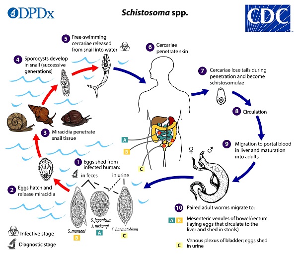
Image: S. haematobium lifecycle (Source: CDC)
Pathogenesis of Schistosoma haematobium
The eggs and cercariae of Schistosoma haematobium are pathogenic while the adult parasites are rarely so.
Pathogenicity due to cercariae
When cercariae penetrate the skin, at the site, it produces allergic dermatitis. Within two hours of infection, a papular pruritic rash appears in hosts with prior exposure/sensitization.
This condition is also seen in individuals infected with avian Schistosoma spp.
Pathogenicity due to eggs
The cause of morbidity and mortality in schistosomiasis is mainly due to the blood fluke’s eggs as it induces a strong inflammatory and granulomatous reaction. This results in the development of granulomas.
The passage of eggs with a terminal spine, from venules into the urinary bladder lumen, causes traumatic lesions which result in hemorrhage. Ectopic lesions such as in the intestine and spinal cord may also occur.
Moreover, due to continuous mechanical irritations and toxins in the eggs, malignant tumors in the urinary bladder may develop.
Pathology of Schistosoma haematobium
Egg granuloma
delayed hypersensitivity reaction around the eggs
found in the ureter, urinary bladder
diameter may vary from 500μm or more
if they join together, even larger nodules are formed
granuloma mainly has Schistosoma haematobium surrounded by T-cells. Eosinophils, macrophages
hyperplastic mucosa shows glandular metaplasia producing cystitis glandularis and it surrounds the nodules
in chronic cases of schistosomiasis, the parasite eggs (calcified or degenerated) lie at the center and are surrounded by fibroblast proliferation and fibrosis
due to fibrosis, marked cicatrization occurs which leads to obstructive uropathies
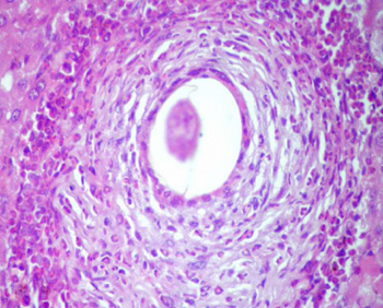
Image: S. haematobium - an active hepatic schistosomal granuloma with a central ovum (Source: ResearchGate)
Host Immunity of Schistosoma haematobium
Concomitant immunity develops in schistosomiasis. This type of immunity protects the infected host from Schistosoma cercariae re-infection but at the same time keeps the adult Schistosoma haematobium alive in the host. The parasite protects itself by antigenic mimicry- coating its teguments with host proteins.
Since eggs of Schistosoma haematobium are highly antigenic, they indicate vigorous host immune response while adult worms and cercarial are extensively less immunogenic.
Moreover, cell-mediated immunity develops numerous types of pathological lesions.