Trichomonas vaginalis - Clinical manifestations, Laboratory diagnosis
Clinical manifestations of Trichomonas vaginalis
The flagellate Trichomonas vaginalis causes a sexually transmitted disease called trichomoniasis or trichomonosis which is a clinical manifestation of the bacteria. The infections are mostly asymptomatic in men than in women. The prognosis is good as metronidazole treats 95% of infections.
In women
Trichomonal vaginitis (Vulvo-vaginitis)
the incubation period for Trichomonas vaginalis is 5-10 days
may become severe after menstruation
the acute stage of Trichomonal vaginitis (Vulvo-vaginitis) may last from a week to a month
in more than 50% of cases, this condition is asymptomatic
symptoms of acute infection include purulent abnormal vaginal discharge, vulvovaginal irritation (50% cases), vaginal erythema, strawberry cervix, etc
the purulent abnormal vaginal discharge, which is seen in two-thirds of cases, includes profuse, purulent yellowish or yellow-greenish discharge which is malodorous
on closer examination of the vagina and cervix by colposcopy mucosa lining the vagina and ectocervix shows punctuate hemorrhage called strawberry mucosa or colpitis macularis and are seen in 45% of cases
other symptoms for Trichomonas vaginalis include abdominal pain, dyspareunia (30%-50% cases), dysuria, disagreeable odor (10% cases)
Urethritis
Urethritis may acquire in a newborn female during vaginal birth and manifests as vaginal discharge during the first week of life
in adult women, this condition may manifest as dysuria and involves the increased frequency of micturition
In Males
more asymptomatic in males than in female
the average incubation period is 10 days but may range from 4 to 28 days
in symptomatic cases, Trichomonas vaginalis causes non-gonococcal urethritis and in rare cases may cause epididymitis, prostatitis, mucopurulent discharge, and at prepuce superficial penile ulcerations can occur
also characterized by pain in the urethra and testicles.
Complications
Trichomonas vaginalis can cause pelvic inflammatory disease (PID) in females
premature rupture of membranes, premature birth, and low birth weight of the baby in pregnant women
in men complications of trichomonosis includes prostatitis, epididymitis, urethral stricture, and infertility
Laboratory diagnosis of Trichomonas vaginalis
Trichomonas vaginalis has been isolated from the cervix, vagina, bladder, urethra, Bartholin, and Skene glands in females.
In males, the parasite has been retrieved from external genitalia, prostate, anterior urethra, epididymis, and semen.
Specimen
from women
vaginal discharge
endocervical swabs (not for wet mount due to the limited number of parasites but used for culture)
urethral discharge
from men
semen
urethral discharge
prostatic fluid
urethral swab before voiding urine in the morning obtained by platinum loop
urine sediment collected during the first early morning void
Microscopy
Wet mount technique
due to the absence of a cyst, iodine wet mount is not done- only normal saline is used
the Trichomonas vaginalis trophozoites are identified by their jerky and twitching motility
a large number of polymorphonuclear (PMN) cells and squamous epithelial cells are also observed which is directly associated with the severity of infection
the trophozoites may not be visible in asymptomatic or chronic cases of infection due to the low number of the parasite
Staining methods
the sample smear can be stained by acridine-orange or Papanicolaou techniques to detect the trophozoites and the sensitivity is similar to wet mount examination
direct fluorescent antibody (DFA) staining can be used in sample smears which is more sensitive than wet mount examination but requires a fluorescent microscope
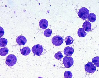
Image: T. vaginalis microscopy (Source: Wikipedia)
Culture
the gold standard for diagnosis of Trichomonas vaginalis with a sensitivity of 95%
for routine tests, the samples are aerobically cultured in-vitro in culture media such as Diamond, Lash, and Kupferberge medium
after 48 hours of incubation at 37° C, motile trophozoites can be observed
usually done in cases where the went mount is negative but the patient shows strong symptoms, resistant trichomoniasis despite treatment, and in high-risk individuals such as sex-workers
Antigen detection
Antigen detection tests such as ELISA can be used to detect antigens of Trichomonas vaginalis in specimens using monoclonal antibodies specific for 65 kDa surface polypeptide.
Serodiagnosis
Serodiagnosis has limited use due to low sensitivity and non-specific reactions.
Molecular diagnosis
The molecular diagnosis of Trichomonas vaginalis includes a DNA probe and PCR.
DNA probe
use of synthetic oligonucleotide probes have been used
high sensitivity and high specificity than the wet mount method
PCR
high sensitivity (97%) and high specificity (98%)