Tuberculosis - Introduction, Virulence Factors, Pathogenesis
Introduction to tuberculosis
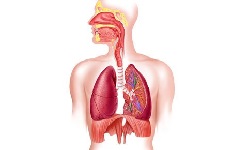
Tuberculosis (TB) is a highly infectious disease caused by Mycobacterium tuberculosis. It typically attacks the lungs, but can also affect other body parts.
Mycobacterium tuberculosis is an acid-fast, intracellular, straight, or slightly curved rod measuring 3x3 micrometers. It occurs in pairs, singles, or as small clumps
The infectious disease is spread through the air when people have active tuberculosis. The risk factors for tuberculosis include- age, immune state, smoking habits, etc.
Most infections are asymptomatic and latent, but about 1 in 10 latent infections eventually progress to active disease if left untreated.
Virulence factors of tuberculosis
The virulence factors of tuberculosis are as follows:
Mechanism of entry: The tubercle bacilli can bind directly to mannose receptors on macrophages
Intracellular growth: Antibodies and complements are not effective as the bacilli grow intracellularly. The bacilli also inhibit phagosome lysosome formation
Slow generation time: Because of the bacilli’s slow generation time, the immune system might not be triggered sufficiently to eliminate them
High cell wall lipid concentration: This accounts for impermeability and resistance to antimicrobial agents. It also provides resistance to killing by acidic and alkaline compounds in both intracellular and extra-cellular environments.
Cord factor: produces serpentine cords found on the surface of the fluid, solid media
Sulpholipids: prevents fusion of phagosome and lysosome inside the macrophage, allowing multiplication inside the macrophages.
Pathogenesis of tuberculosis
Mycobacterium tuberculosis causes tuberculosis. The capability of Mycobacterium tuberculosis for intracellular growth in alveolar macrophages is the main determinant of the virulence of the bacteria. It is a classic representative of an intracellular pathogen.
In primary tuberculosis, the mode of infection starts by direct inhalation of aerosolized bacilli (1-400 in no.) contained in droplets of expectorated sputum, coughing, and sneezing (even a single viable bacillus can lead to infection).
Although the majority of inhaled bacilli are arrested by natural host defense present in the upper respiratory tract (URT), the tubercle bacilli are carried to an alveolus in a droplet nucleus and are phagocytized by alveolar macrophages.
The bacilli are carried to the nearest lymph node, usually in hilar or other mediastinal chains. In the lymph node, the organism multiplies slowly within macrophages resulting in the destruction of infected macrophage cells.
Mycobacterium tuberculosis can multiply inside macrophages either by resistance to lysosomal content or by escape from the phagosome into the macrophage’s cytoplasm inhibiting phagolysosome formation. Tubercle bacilli multiply to a critical mass within the protection from accomplishing phagosome-lysosome fusion capable of destroying the bacteria.
After reaching a critical mass, the Mycobacterium tuberculosis spill out of the destroyed macrophages, through the lymphatics, and into the bloodstream producing mycobacteria and carrying tubercle bacilli to many parts of the body.
In most cases, the host immune system is able to kill these tubercle bacilli. However, a small reservoir of live bacteria may be left in areas of normally high oxygen concentration such as an apical (top) portion of the lungs. These bacilli are walled off.
Those macrophages that kill bacteria, present Ag with MHCII proteins which activate CD4 cells. CD4 cells further, with the help of cytokines, attract macrophages at the site. The macrophages produce lytic enzymes that kill bacilli as well as nearby tissues.
Macrophages fuse to form a giant cell called granuloma (aka hard tubercle) which is more potent. It damages nearby tissue, thus extending cellular damage. If granuloma is not formed, caseous material (solid or semisolid) is left at the primary lesion
In the majority of cases, the lesions heal by themselves leaving a calcified nodule. The Mycobacterium tuberculosis remains in the latent phase, which can reactivate later Inability to eliminate infection can lead to systemic hypersensitivity to Mycobacterium antigens.
Years later an insult to the host, either immunological or physical, may cause the breakdown of the focus of latent tubercle bacilli, allowing active multiplication and disease i.e. secondary tuberculosis.
In cases of immunocompromised host, initial bacteremia seeds tubercle bacilli throughout the host, leading to disseminated or miliary tuberculosis.
Growth of bacilli within host macrophages and histiocytes in the lungs causes an influx of more effector cells, including lymphocytes, neutrophils, and histiocytes, eventually resulting in granuloma formation, then tissue destruction and cavity formation.
The Mycobacterium tuberculosis infection can extend into bronchioles and bronchi from which bacteria are disseminated via respiratory secretions and coughing. Aerosolized droplets are produced by coughing and contain organisms that when inhaled by a susceptible host produce tuberculosis disease.


.jpg)