Babesia - Laboratory Diagnosis, Treatment, Prevention, Control
Laboratory Diagnosis of Babesia
The laboratory diagnosis of Babesia spp depends on the demonstration of intra-erythrocytic parasites in the peripheral blood samples. A patient with a history of tick bites or exposure to ticks in a febrile stage can be suspected of babesiosis.
Specimen
Blood
* Blood samples/specimen should be collected before starting treatment. The collection of blood samples is done from peripheral areas such as the earlobe, finger, and from the greater toe in infants during the time of fever.
Microscopy
The laboratory diagnosis of Babesia spp depends on the microscopic demonstration of intra-erythrocytic parasites in the peripheral blood samples. Multiple peripheral-stained thin smears are examined before ruling out Babesia infection.
Thin blood smear
in thin blood smear method, the thin blood smears are air-dried rapidly, fixed with alcohol, and stained
the tail end of the smear is examined under oil-immersion
Leishman, Wright, or Giemsa stain on thin blood smears is done
Like Plasmodium species, Babesia measures 2 μm to 3 μm with red-blue cytoplasm and red-stained nucleus which is often confused with P. falciparum.
Microscopy of Babesia spp is different from Plasmodium with the following features:
brown pigment deposits (i.e. hemozoin) are absent
gametocytes are absent
merozoites are arranged in Maltese cross forms or pathognomonic tetrads
in cases of heavy infections, trophozoites are seen outside erythrocytes
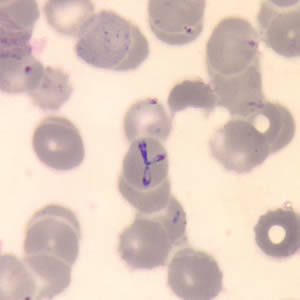
Image: Babesia microscopy after thin Giemsa stain (Image: CDC)
Culture
The culture of Babesia can be done in:
In Media
Babesia bovis can be cultured in the tissue culture medium I99 which has been supplemented with bovine serum
Laboratory animals
Babesia microti can be isolated in hamsters and gerbils
1 ml of EDTA-whole blood sample is intra-peritoneally inoculated in golden hamster and gerbil
the parasite can be demonstrated in the peripheral blood 2-4 weeks after inoculation of the test specimen
Serodiagnosis
in the serodiagnosis of B. microti infection, the indirect fluorescent antibody (IFA) test utilizes intra-erythrocytic Babesia as an antigen
IFA titer of 1:256 or higher is diagnostic for B. microti infection while titers of 1:32 or less indicate past infection
IFA has less specificity due to cross-reaction with sera from malaria infection
Molecular diagnosis
A molecular diagnosis such as PCR is used in the diagnosis of very low levels of parasitemia. It is also useful in patients who previously had babesiosis and have recrudescent Babesia.
Treatment of Babesia
Babesia microti infection in humans is mostly self-limiting and in otherwise healthy patients, recovery occurs without specific chemotherapy.
Common drugs used for the treatment of Babesia include Clindamycin and oral quinines for 7-10 days.
A combination of atovaquone and azithromycin is used as an alternative therapy.
Chemotherapy is not successful in cases of babesiosis.
Prevention, Control of Babesia
Prevention, Control of Babesia includes:
avoid exposure to ticks
use of tick repellants
avoid areas endemic to babesiosis by immuno-compromised or splenectomized individuals
use of proper clothing to prevent a tick bite