Fasciolopsis buski - Life Cycle, Pathogenesis, Pathology, Host Immunity, Laboratory Diagnosis, Treatment
Life Cycle of Fasciolopsis buski
The life cycle of Fasciolopsis buski occurs in the following hosts while aquatic vegetations are also mandatory.
Definitive host: Pig and Man are the primary definite host.
Intermediate host:
First intermediate host: Planorbid snails of the genera Segmentina, Hippeutis, and Polypylis.
Second Intermediate host: Freshwater plants such as water caltrops (Trapa bispinosa), water hyacinth (Eichlornia), water chestnut (Eleocharis tuberosa), lotus (Nelumbium), water bamboo (Zizania aquatica) harbour the infective stage (metacercariae) of Fasciolopsis buski.
the definitive host acquires infection after consumption of the second intermediate host - water caltrops (Trapa bispinosa), water hyacinth (Eichlornia), water chestnut (Eleocharis tuberosa), lotus (Nelumbium), water bamboo (Zizania aquatica) harbour the infective stage (metacercariae)
once metacercariae reach the duodenum, it excysts where they develop and sexually mature in three months
after mating, they began laying unembryonated eggs
these eggs are passed along with the host faeces
once in the water, further development of Fasciolopsis buski eggs takes place
within 3 to 5 weeks, the egg hatches to release one miracidium larva per egg
the miracidium larvae of the giant fluke must find and invade the planorbid snails of the genera Segmentina, Hippeutis, and Polypylis – otherwise, it dies
once inside its primary intermediate host, the miracidium larvae underdo asexual reproduction by a single generation of sporocysts, two generations of rediae (mother rediae and daughter rediae)
within a couple of weeks, a single miracidium larva gives rise to a large number of cercariae
the Fasciolopsis buski cercariae then escape from the snail into the water, encyst and adhere to the second intermediate host
after a few hours on the second intermediate host, the cercariae encyst to form metacercariae
if the second intermediate host harbouring metacercariae is consumed, it causes liver fluke infection in the definitive host including the man – thus the life cycle of Fasciolopsis buski is continued
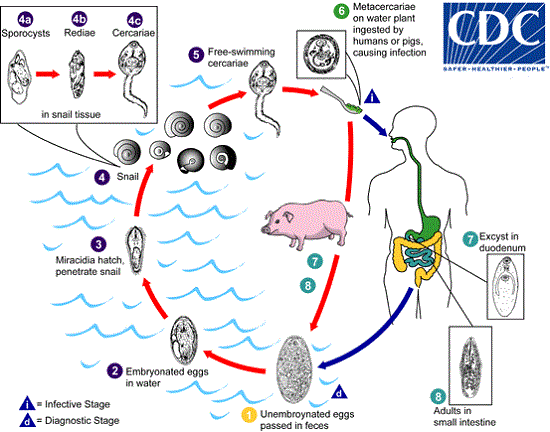
Image: Fasciolopsis buski Lifecycle (Source: CDC)
Pathogenesis, Pathology of Fasciolopsis buski
The key pathological feature of fasciolopsiasis caused by Fasciolopsis buski adults includes traumatic, obstructive, and toxic damage to the intestinal mucosa.
Also, in the duodenum and jejunum, where the adults attach, numerous pathological changes can occur such as localized haemorrhagic inflammation, mucous secretion, and ulceration.
In case of severe infection where a large number of adult flukes are present, the increased mucous secretion results in partial obstruction of the intestinal tract.
This can also result in conditions such as hypoalbuminemia due to malabsorption, impaired B12 absorption, and protein-losing enteropathy. In addition, due to increased metabolite absorption, toxaemia may occur due to toxin formation.
Laboratory diagnosis of Fasciolopsis buski
The laboratory diagnosis of Fasciolopsis buski starts with the collection of samples, followed by microscopy, serodiagnosis, and imaging methods.
Sample
Samples collected include
stool
biopsy tissues (intestine)
blood (serum)
Microscopy
microscopic diagnosis of fasciolopsiasis is commonly done by demonstration of large operculated Fasciolopsis buski eggs
concentrations are done by sedimentation in 0.5% glycerinated saline, formalin-ether concentration method
cannot distinguish between Fasciola hepatica, Fasciolopsis buski and Fasciola buski eggs
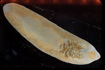
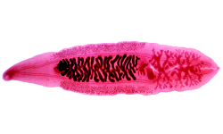
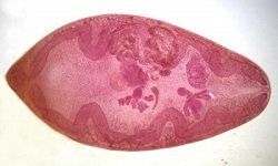
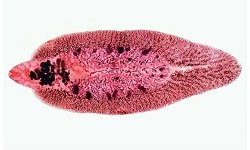
Image: Fasciolopsis buski adult (Source: CDC)
Stool antigen detection
Enzyme-linked immunosorbent assay (ELISA) can be done in stool samples to detect Fasciola corpo-antigen for chronic infection
Molecular test
Important molecular tests of Fasciola hepatica diagnosis include:
DNA probes
PCR
Imaging Methods
Ultrasonogram (USG)
CT-scan
MRI
Treatment of Fasciolopsis buski infection
The drug of choice for the treatment of Fasciolopsis buski infection is Praziquantel.
Prevention, Control of Fasciolopsis buski
The control and prevention of Fasciolopsis buski can be obtained by following steps:
avoid consumption of raw/undercooked aquatic plants
thorough cleaning and decontamination of salad vegetables such as watercress and other water-grown greens
proper filtration and/or boiling of drinking water
immediate treatment of infections in pig
use of molluscicides to control snails
reduce contamination of water sources by pig, and human faeces