Strongyloides stercoralis - Introduction, Classification, History, Habitat, Morphology
Introduction to Strongyloides stercoralis
Strongyloides stercoralis, commonly known as the dwarf threadworm, is the causative agent of strongyloidiasis. The parasitic nematode is infectious to humans and has a unique feature – both parasitic and free-living generations.
Some of the characteristic features of Strongyloides stercoralis include:
the parasite can develop into a free-living generation in the soil i.e. outside the human host
while travelling in the host intestine the larval stage of the parasite might develop into filarial larvae
since males are absent, the females reproduce parthenogenically
eggs containing mature larvae are released by the female
History of Strongyloides stercoralis
Historically, in 1876, Normand was the first to demonstrate Strongyloides stercoralis in the stool samples collected from French soldiers suffering from diarrhoea. In 1928, Nishigori discovered autoinfection by Strongyloides stercoralis present in the host bowel by the rhabditiform larvae.
Habitat of Strongyloides stercoralis
The female Strongyloides stercoralis parasite habitats the mucosa of the small intestine – especially the duodenum and upper jejunum. They lie in the tunnels of the enterocytes of the intestinal mucosa.
Morphology of Strongyloides stercoralis
The morphological stages of Strongyloides stercoralis include – parasitic adult worms (male, female), eggs, free-living worms (male, female), and larvae (rhabditiform larvae, filariform larvae).
Adult worm
The adult worms of the parasite include the parasitic male and the parasitic female
Parasitic male
parasitic males of Strongyloides stercoralis have not been found to be infectious to humans
however, the male parasite has survived in experimentally infected dogs
shorter and broader than females
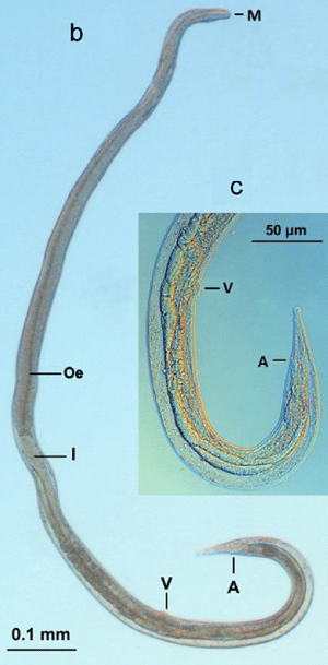
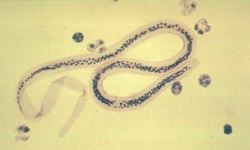
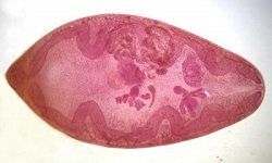
Image: parasitic female Strongyloides stercoralis - (A) Anus, (GP) genital primordium, (I) intestine, (M) mouth, (Oe) oesophagus, (V) vulva (Source: ResearchGate)
Parasitic female
parasitic females of Strongyloides stercoralis are small
measures 2.5 mm in length and 0.4 mm to 0.5 mm in breadth
translucent
four small lips in the buccal cavity
the anterior end has a long, cylindrical oesophagus
the posterior two-third part of the body contains the intestine
the anal opening is present in the mid-ventral area, a little far away from the caudal tip
genital organs include a pair of uteri, oviduct, and ovaries
at the junction of the middle and posterior ends of the body, the vulval opening is present
ovo-viviparus
each parasite can lay 30-40 partially embryonated eggs per day
the eggs are released in the mucosal epithelium of the intestine
lifespan is nearly a year
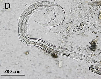
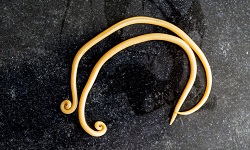
Image: free-living Strongyloides stercoralis male (Source: ajtmh)
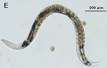
Image: free-living female Strongyloides stercoralis morphology (Source: ajtmh)
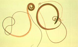
Free-living male and female
The free-living form of the male and female Strongyloides stercoralis measure 0.7 mm by 1 mm in size. They live in their natural habitat – the soil and multiply sexually.
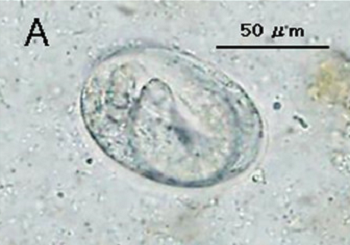
Image: Strongyloides stercoralis embryonated eggs (Source: ajtmh)
Eggs
eggs of Strongyloides stercoralis are similar to that of hookworm but are smaller in size
measures 55 μm to 60 μm in length and 30 μm to 35 μm in breadth
oval, transparent, and thin-shelled
each contains larvae which are ready to hatch
immediately after eggs are laying, the rhabditiform larvae hatch and migrate back to the intestinal lumen
passed out along with the host faeces
due to quick hatching, only larvae are found in faeces but not in the faeces
Larva
The larva of Strongyloides stercoralis consists of two stages - rhabditiform larvae, and filariform larvae.
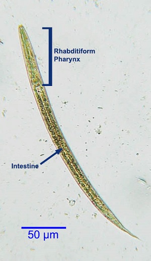
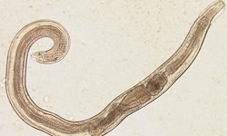
Image: Strongyloides stercoralis rhabditiform larvae (Source: Western College of Veterinary Disease)
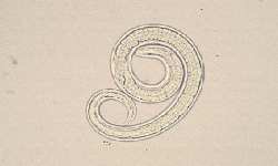
Rhabditiform larvae
rhabditiform larvae of Strongyloides stercoralis are the first-stage larvae of Strongyloides stercoralis
immediately hatch out of eggs after being laid by the gravid female
found in the faeces
motile and unsheathed
measures 200 μm to 300 μm in length and 16 μm in breadth
has a short mouth and double-bulb oesophagus
has an inconspicuous genital primordium
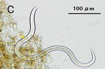
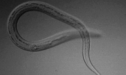
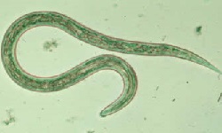
Image: Strongyloides stercoralis filariform larvae (Source: ajtmh)
Filariform larvae
filariform larvae of Strongyloides stercoralis are in the infective stage of humans
delicate body with a short mouth and long cylindrical oesophagus
measure 630 μm in length and 10 μm in breadth
cause infection by penetrating the host skin