Schistosoma japonicum - Laboratory Diagnosis, Treatment, Prevention, Control
Laboratory diagnosis of Schistosoma japonicum
The laboratory diagnosis of Schistosoma japonicum starts with the collection of samples, followed by microscopy, serodiagnosis, and imaging methods.
Diagnosis of schistosomiasis is commonly done by demonstration of typical non-operculate lateral-spined eggs.
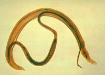
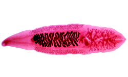
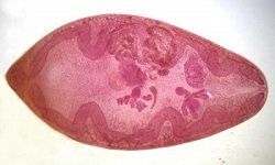
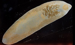
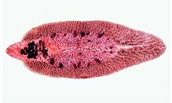
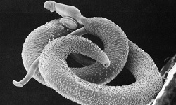
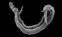
Image: Schistosoma japonicum adults (Source: ruby.fgcu.edu)
Samples
stool
blood (serum)
autopsy/biopsy (intestine, liver)
CSF
Bronchoalveolar lavage (BAL)
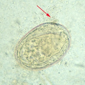
Image: Schistosoma japonicum egg with a central spine in a depression (red arrow) (Source: CDC)
Microscopy
Microscopy of stool, biopsy, and autopsy samples is the most common method as in acute cases, Schistosoma japonicum eggs can be demonstrated by direct faecal smear microscopy.
In chronic cases, the number of eggs released in the faeces can be scanty and intermittent, concentrations are done by sedimentation in 0.5% glycerinated saline, acid-ether method.
For quantitative diagnosis, Katos’ cellophane faecal thick smear is performed to assess the degree of infection as well as treatment response.
If active infection is to be tested, a hatching test with Schistosoma japonicum eggs is carried out. In this test fresh stool specimen is checked for newly hatched miracidium larvae.
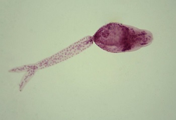
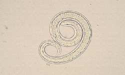
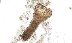
Image: Schistosoma japonicum cercaria (Source: CDC)
Detection of antibodies
Serodiagnosis methods can be used to demonstrate specific circulating antibodies in the patient's serum for Schistosoma japonicum. It uses purified schistosome antigens to antigen fractions with specific antigenic determinants.
These tests are mostly used to detect infection during the pre-patent period, chronic infection, and ectopic infection as well as for epidemiological studies.
However, its major disadvantage is its inability to differentiate between active and past infections as antibodies remain in the serum for a long time. Also, these tests cannot quantify egg/infection load.
Schistosoma-specific antibody detection methods include:
Indirect haemagglutination (IHA)
Radio Immuno Assay (RIA)
enzyme-linked immunosorbent assay (ELISA)
Falcon assay screening test (FAST)-ELISA
* FAST has high sensitivity (95%) and specificity (99%)
* uses haematobium adult worm microsomal antigen (HAMA)
Immunoblot
Detection of antigen
These tests are based on the detection of Schistosoma japonicum antigen in the sample. They also help in the detection of autoinfection and the difference between active and past infections.
The tests include:
immunoelectrophoresis-in-gel, counter-current immunoelectrophoresis (CIEP)
enzyme-linked immunosorbent assay (ELISA) – high sensitivity
* demonstration of proteoglycan gut-associated antigens such as circulating anodic antigen (CAA) and circulating cathodic antigen (CCA)
Radio Immuno Assay (RIA)
Imaging methods
X-ray
Ultrasonography (US) if the liver, and spleen are involved
CT scan (if CNS is involved)
MRI (if CNS is involved)
Other tests
Histological test (rectal biopsy) for intestinal schistosomiasis
Treatment of Schistosoma japonicum
Praziquantel is the drug of choice because Schistosoma japonicum is resistant to most treatments
Prevention, Control of Schistosoma japonicum
immediate treatment of infected definitive hosts
use of molluscicides to control snails
education and awareness
reduce contamination of water sources by human urine