Trichophyton rubrum - Laboratory diagnosis
Laboratory diagnosis of Trichophyton rubrum
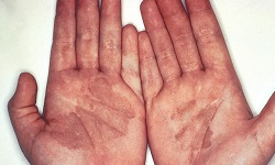
Laboratory diagnosis of Trichophyton rubrum is done by demonstration of fungal particles such as hyphae, and spores in the specimen.
Samples
Hair
Skin
Nail scrapping
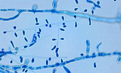
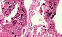
Microscopy
Microscopy (direct microscopy) is performed following KOH preparation to demonstrate fungal particles in the specimen.
KOH preparation of Trichophyton rubrum under microscope (Source: ResearchGate)
KOH preparation
The procedure of KOH preparation for fungal infection diagnosis is as follows.
Procedure
the specimen is placed on a clean, grease-free slide and a drop of 10-20% KOH solution is added to it
the sample is covered with a coverslip and immediately viewed under a microscope
the KOH preparation is examined again after 20 mins
in skin or nail specimens, chains of spores or branching hyphae are seen
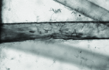
in hair specimen, parallel rows of spores are observed outside (ectothrix) or inside (endothrix) the hair shaft
* infected hair shaft may also contain air spaces
* skin test does not distinguish active infection
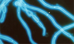
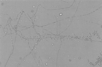
Hair perforation test showing erosion by Trichophyton rubrum (Source: ResearchGate)
Hair perforation test
The hair perforation test is done mostly to distinguish Trichophyton rubrum from Trichophyton mentagrophytes, which are morphologically similar.
Trichophyton rubrum causes only surface erosion of the hair shaft while from Trichophyton mentagrophytes infection results in wedge-shaped perforation.
Procedure
hair specimens (5-10mm in size) are placed in a Petri dish with 20ml distilled water and autoclaved
sterile 10% yeast extract (2-3 drops) is added to the Petri dish
the hair strands are inoculated with small fragments of test fungi that have been grown on SDA
the inoculated hair strands are incubated at 25°C
for up to a month, the inoculated hair strands are examined weekly after the addition of Lacto Phenol Cotton Blue (LPCB)
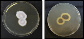
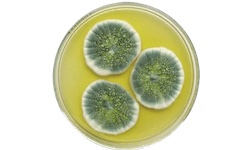
Trichophyton rubrum frontside (left) and reverse side (right) on SDA (Source: ResearchGate)
Culture
The culture of Trichophyton rubrum can be done as a diagnostic step.
The specimen is inoculated onto Sabouraud’s Dextrose Agar (SDA) incorporated with chloramphenicol and cycloheximide and incubated at 25 to 30°C aerobically for 1 to 3 weeks.
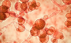
The fungi are identified on the basis of colony morphology, color, pigment production, and the presence/absence of microconidia or macroconidia.
Fresh culture of extracted from the agar plate using cellophane tape and placed on a slide containing a drop of Lacto Phenol Cotton Blue (LPCB). The preparation is viewed under a microscope to demonstrate the presence/absence of microconidia or macroconidia.
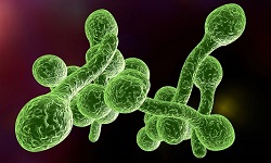
Trichophyton rubrum colonies are cottony which later turns velvety with red pigment on the reverse side. Numerous microconidia are numerous, teardrop-shaped, and present along the side of the hyphae. Microconidia are usually absent but if present are few in number, smooth, thin-walled, and pencil-shaped.
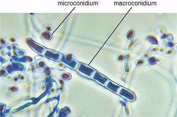
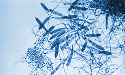
Trichophyton rubrum microconidia, macroconidia (Source: Creative biolabs)
Urease test
A urease test is done for distinguishing Trichophyton rubrum (urease negative) from Trichophyton mentagrophytes (urease positive).
Procedure
a sterile Christensen’s urea agar is taken and inoculated with test fungi
the test tube is incubated at 25°C for 5 days
if the color of the agar changes to pink in color, it is urease positive
Molecular test
The molecular tests used for the diagnosis of Trichophyton rubrum are:
PCR-reverse line blot test
real-time PCR test
multiplex PCR test
PCR-ELISA test
MALDI-TOF test