Trypanosoma cruzi - Laboratory diagnosis, Prevention, Treatment, Control
Laboratory diagnosis of Trypanosoma cruzi
The laboratory diagnosis of Trypanosoma cruzi beings by collection of samples:
Samples
The specimens for lab diagnosis of Trypanosoma cruzi include:
blood
tissue biopsy
tissue aspiration
Microscopy
Wet mount
demonstration of mobile Trypanosoma cruzi trypomastigotes in a direct wet mount of anticoagulated sample blood microscopy
the presence of a buffy coat during microscopy
a hundred microscopic fields must be done before concluding the sample is negative for Changa’s disease
Blood smears
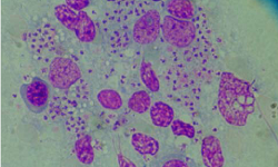
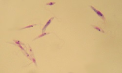
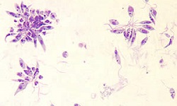
both thick and thin blood smear microscopy stained by Giemsa stain is done
less sensitive procedure as the Trypanosoma cruzi parasites are easily disrupted during the preparation of the smear
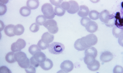
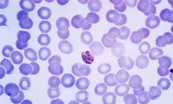
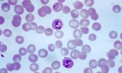
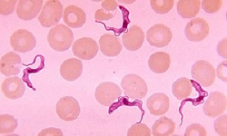
the main advantage is that the Giemsa stain thin blood smear stains the kinetoplast and flagella of the trypomastigotes which helps in the identification and differentiation of Trypanosoma cruzi from Trypanosoma rangelli
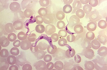
Figure: Trypanosoma cruzi Giemsa stain microscopy (Source: CDC)
Concentrated blood microscopy
Microhaematocrit and Strout’s methods are used for the concentration of blood samples for examination of the Trypanosoma cruzi parasite.
In the Microhaematocrit method
75μl of blood sample is placed in a heparinized capillary tube
centrifuged for 5 mins to 10 minutes
wet mount of the buffy coat is observed under a microscope for motile trypomastigotes
In Strout’s method
3 ml of sample blood is incubated at 37° C for an hour
collection of serum and centrifugation at 100g for 3 minutes
the supernatant is collected and centrifuged at 400g for 5 mins
the final precipitate is examined under a microscope for Trypanosoma cruzi trypomastigotes
Culture
The culture of Trypanosoma cruzi is done via blood culture and via animal inoculation.
Blood culture
blood culture methods are followed if repeated microscopy failed
it is done in NNN media or liver infusion tryptose (LIT) medium
incubated at 22° C - 24° C for four to six months
the culture is examined by the 4th day and every week for 6 months
presence of epimastigotes and trypomastigotes confirms Trypanosoma cruzi infection
Animal inoculation
samples are inoculated intraperitoneally into laboratory mice
tail blood is collected for 10 days and examined microscopically for motile trypomastigotes
two months after inoculation, the mice are killed and heart tissues are examined for the parasite
Xenodiagnosis
Xenodiagnosis is used for the diagnosis of acute as well as chronic Chagas’ disease
done by feeding the patient 14-20 instar nymph (3rd or 4th-day larvae) of reduviid bug for 3 consecutive days
the bugs are then maintained laboratory and their faces are examined once a month for Trypanosoma cruzi amastigotes, epimastigotes, and trypomastigotes
in chronic cases, the sensitivity is 50% for this method
Serodiagnosis
commonly used serological methods for Trypanosoma cruzi diagnosis include indirect haemagglutination (IHA), Indirect immunofluorescent antibody (IFA), Direct Agglutination test (DAT), Enzyme-Linked Immunosorbent Assay (ELISA)
limited value due to cross-reactivity with leishmaniasis and syphilis
mostly used in screening in pregnant women, blood donors, seroepidemiological studies, assessment of asymptomatic carriers
Molecular methods
The molecular methods of Trypanosoma cruzi are as follows:
PCR
DNA probes
Other tests
Chest radiography shows an enlargement of the heart in acute or chronic cases
ultrasound detects intracardiac thrombi, hypokinesia of the septal cardiac walls, or ventricular dysfunctions
ECG detects cardiomyopathy
esophageal endoscopy visualized megaoesophagus
Treatment of Trypanosoma cruzi
Treatment of Trypanosoma cruzi can be done by:
Chemotherapy is unsatisfactory
drugs used include Nitfurtimox and benznidazole
the drugs only kill extracellular parasites but not intra-cellular parasites
Gentian violet can prevent the transmission of parasites via blood transfusion
Prevention, Control of Trypanosoma cruzi
The prevention, control of Trypanosoma cruzi can be achieved by the following:
reduction of the reduviid bug population by using insecticides
evade contact with potential animal vectors such as dogs, rodents, or other mammals
use of bed nets, window nets, or insect repellents
Screening of blood donors