Wuchereria bancrofti - Introduction, Classification, History, Habitat, Morphology
Introduction to Wuchereria bancrofti
Wuchereria bancrofti, also known as Bancroft’s filarial worm, is the etiological agent of lymphatic filariasis. The lymphatic filariasis caused by the filarial worm Wuchereria bancrofti is also called bancroftian filariasis.
Classification of Wuchereria bancrofti
The classification of Wuchereria bancrofti is as follows:
Kingdom: Animalia
Phylum: Nematoda
Class: Chromadorea
Order: Rhabditida
Family: Onchocercidae
Genus: Wuchereria
Species: W. bancrofti
History of Wuchereria bancrofti
In 1862, Jean-Nicolas Demarquay was the first one to demonstrate microfilariae in hydrocele fluid from a man from Havana, Cuba. Otto Wucherer also found microfilariae in the urine of a woman with chyluria in Bahia, Brazil in 1866.
Unaware of both previous demonstrations, Timothy Lewis found Wuchereria bancrofti microfilariae in the urine of an Indian with chyluria in India in 1870. He again reported observation of the parasites in the blood just two years later.
Joseph Bancroft in Brisbane, Australia discovered adult filarial parasites in the lymphatic abscesses in patients with larvae in the blood in the years 1876 and 1877. After Bancroft sent the specimens to London, Spencer Cobbold named them Filaria Bancrofti.
In Xiamen, China, in 1877, Patrick Manson discovered if Culex quinquefasciatus and Aedes aegypti mosquitoes took a blood meal from a person with larvae (microfilariae) in the blood, the parasites molted twice in the insects' abdomen to become larger worms. In 1879, he discovered that blood-dwelling forms of Wuchereria bancrofti had a nocturnal periodicity and appeared in the host blood in large numbers around midnight while during the day, minimal numbers are seen in blood.
Thomas Bancroft in 1899 let laboratory-reared mosquitoes feed on a patient infected with microfilaraemia. He then kept the infected arthropods for 16 days and also sent some specimens to George Low in London.
In London, Low prepared histological sections of the mosquitoes and discovered that the Wuchereria bancrofti larvae had migrated from the abdomen of the mosquito to the thorax to salivary glands, and then to the proboscis.
Thomas Bancroft in 1902 demonstrated the mode of transmission for Wuchereria bancrofti is via the bite of an infected mosquito by using Dirofilaria immitis, a related worm. He showed adult worms in experimentally infected dogs.
In 1921, Léon Seurat made the genus Wuchereria and placed the filarial nematode parasite in it.
Habitat of Wuchereria bancrofti
The adult Wuchereria bancrofti parasites are found in the lymphatic vessels – especially the lymph nodes of humans and other vertebrates.
The larval form, microfilaria, is the first stage larvae which are found in the peripheral blood. In some cases, they are also found in chylous urine or in hydrocele fluid.
Morphology of Wuchereria bancrofti
The significant morphological forms of Wuchereria bancrofti include adult, microfilaria (first-stage larvae), and third-stage larvae.
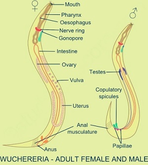
Wuchereria bancrofti morphology (Source: Lab Weeks)
Adult form
adult worms of Wuchereria bancrofti are whitish, minute, and thread-like
filariform in shape with a smooth surface
both anterior and posterior ends are tapering
the anterior end is slightly swollen with two rows of ten sessile papillae surmounted
a posterior end consists of the anus in its terminal end
obtain nutrients for growth from the lymph of the lymphatic system
the average lifespan is around 4 years to 5 years
Male
male Wuchereria bancrofti measures 40 mm in length and 0.1 mm in diameter
sharply curved ventral tail with two spicules of unequal length
Female
female Wuchereria bancrofti is ovoviviparous
longer than males
length may range from 80 mm to 100 mm and 0.24, to 0.30 mm in diameter
have curved tail
lays eggs containing well-developed embryos i.e. microfilariae
in a gravid female, the microfilariae remain coiled together in the uterus and vagina
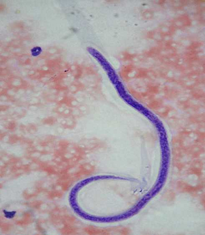
Wuchereria bancrofti microfilaria (Source: ResearchGate)
Microfilaria
microfilaria of Wuchereria bancrofti is called the first-stage larvae
found in the peripheral blood, hydrocele fluid, and chylous urine
size varies from 244 μm to 296 μm in length and 7.5 μm to 10 μm in diameter
are transparent, and colorless with bluntly rounded anterior ends and pointed tails but can be stained to visualize internal structures
covered by a hyaline sheath
has a central column of nuclei with characteristic anatomical landmarks – including nerve ring (nr), excretory pore (ep), excretory cells (ec), anal pores (ap), and gulls
body nuclei are fewer but more distinct and lack nuclei at the end of its tail
can live upto 70 days in the human blood
Infective form
The infective form of Wuchereria bancrofti is the third-stage larvae.
Third-stage larvae
the infective form of Wuchereria bancrofti
found in mosquito vectors
measures 1.4 to 2 μm in length and 18 to 23 μm in breadth
elongated and filariform of the parasite