Echinococcus granulosus - Life Cycle, Pathogenesis, Pathology, Host Immunity
Life Cycle of Echinococcus granulosus
The life cycle of Echinococcus granulosus is completed in two hosts:
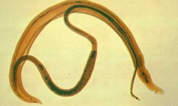
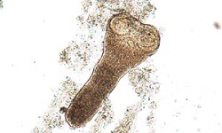
Definitive host: Dog (optimum host) and wild canines such as fox, wolf, and jackal. Adult tapeworms live in the intestine and release a large number of eggs along with host feces.
Intermediate host: Man (accidental host), sheep (optimum host), cattle, camel, pigs, horses, and other intermediate hosts which are mostly herbivores. Hydatid cyst i.e. the larval stage is found in these hosts.
intermediate hosts acquire the infection after ingestion of Echinococcus granulosus eggs contaminated in food or water
within eight hours of ingestion, the eggs hatch in the small intestine to release oncospheres or embryos
the embryos penetrate the intestinal wall from where they are carried to various organs (mostly liver and lungs) where they lodge through the body via the bloodstream
if embryos survive the initial inflammatory response of the host, it increases in size up to 5cm - 8cm in a few months
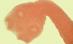
it eventually transforms into a fluid-filled hollow bladder called a hydatid cyst
from a number of nuclei present in the inner germinal layer of the cyst, buds of tissue develop into hollow, fluid-filled capsules known as brood capsules
these brood capsules use their peduncle to attach to the cyst wall or may float free in the hydatid fluid
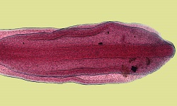
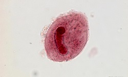
many Echinococcus granulosus larvae (protoscolices) develop from the germinal layer within the brood capsule in large numbers
as cysts grow older and larger, a large number of brood capsules as well as free protoscolices float inside the hydatid fluid called the hydatid sand
these mature cysts (protoscolices) are infectious for the definitive hosts such as dogs
the definitive hosts acquire infection by ingestion of hydatid cysts containing protoscolices present in the viscera of sheep and other intermediate hosts
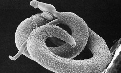
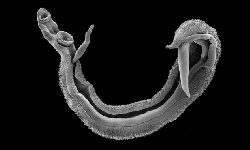
the protoscolices evaginates and penetrate deep between villi and enter the crypts of Liberkuhn in the small intestine
the Echinococcus granulosus develop into an adult tapeworm within 41 days to 76 days after ingestion of hydatid cyst
massive infection in dogs can occur from a single hydatid cyst as it can give rise to thousands and even tens of thousands of adult worms
these parasites release eggs along with the host feces and the life cycle continues
however, if the intermediate host is man, dogs have no access to the infected viscera of man and are the dead end for the parasite
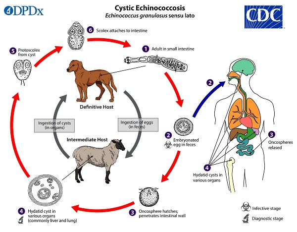
Image: E. granulosus lifecycle (Source: CDC)
Pathogenesis, Pathology of Echinococcus granulosus
Depending on the course of the infection, the fate of Echinococcus granulosus hydatid cysts is different. Some cysts may grow to a certain size and remain so without producing pathological changes for many years while other cysts may rupture/collapse and disappear.
Since Echinococcus granulosus hydatid cysts are responsible for the pathogenesis of the disease, cystic echinococcosis is of two types- primary cystic echinococcosis and secondary cystic echinococcosis.
Primary cystic echinococcosis
Primary cystic echinococcosis occurs after per-oral infection with Echinococcus granulosus eggs which gives rise to hydatid cysts in different parts of the body
the hydatid cysts can be found in any organ but the most common sites are the liver followed by the lungs and other organs
in 80% of cases, the cysts are found only in one organ while in 20% of cases, the cyst might be disseminated to multiple sites and organs
in the organs, the cysts evoke a foreign body type reaction in surrounding tissues but never invade the infiltrates the tissues
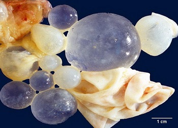
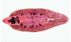
Image: E. granulosus hydatid cyst removed from liver (Source: webpathology)
Secondary cystic echinococcosis
Secondary cystic echinococcosis occurs when primary hydatid cysts rupture as a result of trauma
protoscolices reach different sites via blood circulation where they develop into secondary hydatid cysts
Host Immunity of Echinococcus granulosus
The host immune response in men involves elevated circulating antibodies, particularly the immunoglobulin IgG in the serum. Although these circulating immunoglobulins are parasite-specific they are not protective and do not confer any protection.
Casoni’s intradermal skin test in vivo and transformation of lymphocytes in vitro demonstrates the appearance of delayed hypersensitivity reactions. However, in 10% of cases, the Echinococcus granulosus hydatid disease shows no immune response but the cause of such lack of response is not known.