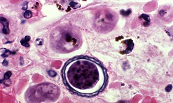
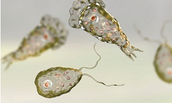
Entamoeba histolytica - Introduction, History, Habitat, Morphology
Introduction of Entamoeba histolytica
The parasitic protozoa Entamoeba histolytica causes the disease amoebiasis. It predominantly infects humans as well as other primates and is worldwide in distribution.
In developing countries, amoebiasis is the third leading cause of parasitic death while in developed countries, Entamoeba histolytica infection is an important causative agent of diarrhea in AIDS patients.
Classification of Entamoeba histolytica
Phenotypically, Entamoeba histolytica is classified as:
Domain: Eukaryota
Phylum: Amoebozoa
Family: Entamoebidae
Genus: Entamoeba
Species: E. histolytica
History of Entamoeba histolytica
W.D. Lambli, in 1859, was the first person to demonstrate the presence of parasitic protozoa in the colon autopsy of a child who had died of diarrhea. In 1875 Fedor Losch first described the parasite. He also proved it was a pathogen by infecting a dog with dysentery stool and showing the produced intestinal lesions.
However, Kartulis, in 1886, was one to conclusively demonstrate that the parasitic amoeba is the causative agent of amoebic dysentery and amoebic liver abscess.
Decades later, in 1913, Walker and Sellards successfully demonstrated that the cyst form of Entamoeba histolytica is the infective form. Only in 1925 did Dobell first describe the life cycle of Entamoeba histolytica.
Habitat of Entamoeba histolytica
Entamoeba histolytica habitats the lumen, mucosa, and submucosa of the large intestine in humans.
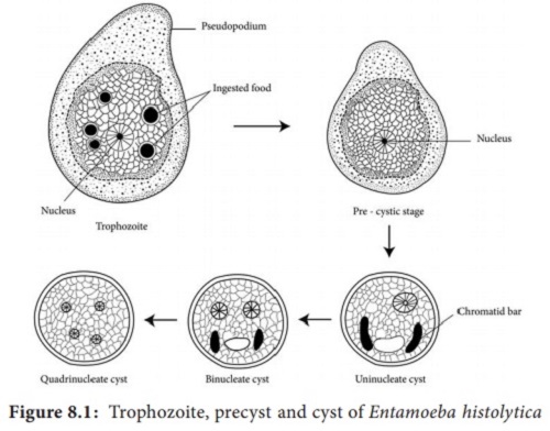
Fig: E. histolytica morphological forms (Source: BrainKart)
Morphology of Entamoeba histolytica
The morphology of Entamoeba histolytica exists in three stages:
Trophozoite
Pre-cyst
Cyst
Trophozoite
This morphological stage of Entamoeba histolytica is the invasive and anaerobic form of the parasitic protozoa
trophozoite size varies from 8μm to 30 μm with an average of 15μm
Its single nucleus is spherical in share, measuring 3.5μm in size
Presence of central karyosome, peripheral chromatin
The cytoplasm contains clear ectoplasm and granular endoplasm which holds food vacuoles ad erythrocytes
The presence of ingested erythrocytes in the endoplasm is a characteristic in the identification of Entamoeba histolytica
Lacks mitochondria, Golgi bodies, and rough endoplasmic reticulum
Motility
The motility of Entamoeba histolytica takes place with the help of Pseudopodia which is a clear hyaline ectoplasm. The motility occurs by the forward extension of the hyaline ectoplasm with granular endoplasm. The mechanism of the flow/extension of ectoplasm and endoplasm produces finger-like extension.
Observation by light microscopy reveals the presence of filopodia.
Pre-cyst
Entamoeba histolytica pre-cyst is smaller than Entamoeba histolytica trophozoite and larger than a cyst
measures 10 μm to 20 μm
round, oval, colorless, and has blunt pseudopodium projecting from the periphery
contains a large glycogen vacuole, two chromatid bars, and rounded the ends
Cyst
this infective form of Entamoeba histolytica which is small and spherical
Entamoeba histolytica cyst is 10 μm to 16 μm in diameter
maturity confirmed by the presence of four nuclei
granular cytoplasm containing rod-like chromatin bars and glycogen mass
lacks red blood cells or food particles
the presence of a thin, smooth chitinous wall around the cyst makes it highly resistant to gastric acid, chlorine in potable water, and other adverse environmental conditions
found in mostly formed stools as well as in semi-formed stools