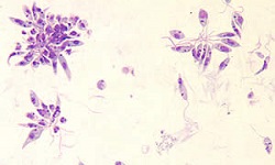
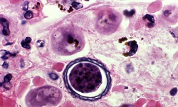
Entamoeba histolytica - Pathology of intestinal, extra intestinal amoebiasis
Pathology of intestinal amoebiasis of Entamoeba histolytica
The pathology of intestinal amoebiasis of Entamoeba histolytica includes:
Amoebic ulcer in the large intestine
Amoebic ulcer in the large intestine is the characteristic lesion, which is flask-shaped, in the intestinal amoebiasis
the site of the Entamoeba histolytica ulcer is associated with the location of the entry in the intestinal mucosa
the localized amoebic ulcers occur in the ileocaecal region- mainly in the caecum and ascending colon and less commonly in the sigmoidal-rectal region while they may also be present in the ileocaecal valve and appendix
the generalized Entamoeba histolytica ulcers are scattered throughout the entire length of the large intestine- even up to the internal anal sphincter
although the initial lesions are small, the diameter of the intestinal ulcer may vary from pinhead to an inch in diameter
lesions may extend only to the muscularis mucosa but sometimes the ulcer penetrates through the mucosal layer and may form a thin membranous base.
the mucous surrounding the Entamoeba histolytica ulcer is slightly oedematous, the ulcer margins are hyperemic while the ulcer is covered with mucus and abundant trophozoites
the base of the ulcer is covered by yellowish and black scars, which are found to be projecting into the intestinal lumen
in case the ulcer progresses, the superficial lesions deepen and can be as big as 1cm in diameter and extend into the sub-mucosal layer forming a broad base and a narrow neck - often described as flask-shaped ulcers
if the Entamoeba histolytica ulcer penetrates through the serosa, peritonitis or pneumo-peritoneum is formed
in case, the ulcer directly extends from the intestine, lesions may appear on the skin of the personal areas of the genital organs- penis in males and vagina, vulva, or cervix in females
if the amoebiasis is acute fulminant, entire mucous membranes may become oedematous and gangrenous which may result in hemorrhage, generalized peritonitis, pericaecal or pericolic abscess, gangrene of the large intestine, and sloughing
in cases of chronic amoebiasis, the ulcers are small and shallow, only extending to the mucosa, but are extensive. This condition is associated with thinning, dilation, and sacculation of the intestinal wall. But in some individuals, generalized thickening of the intestine occurs which causes the narrowing of the intestinal lumen
In rare cases, intestinal amoebiasis caused by Entamoeba histolytica results in the formation of granulomatous tissue called amoebic granuloma – without any ulceration
Pathology of extra-intestinal amoebiasis of Entamoeba histolytica
Pathology of extra-intestinal amoebiasis of Entamoeba histolytica includes amoebiasis of amoebic liver abscess (ALA) which is the most common form
the amoebic pulmonary abscess caused by Entamoeba histolytica is observed less frequently while in rare cases cerebral, cutaneous, and splenic amoebic abscesses also occur
Amoebic liver abscess (ALA)
Amoebic liver abscess (ALA) caused by Entamoeba histolytica may be single or multiple abscesses and can be present in any part of the liver, most commonly in the right lobe’s posterosuperior surface
the ulcer has three zones –
* an inner central necrotic zone containing lysed hepatocytes (no trophozoites)
* intermediate zone with degenerated liver cells, red cells, leucocytes, few trophozoites, and connective tissue cells
* the outer zone consists of normal liver tissue which is being invaded by trophozoites