Schistosoma mansoni - Introduction, Classification, History, Habitat, Morphology
Introduction of Schistosoma mansoni
Schistosoma mansoni is a major blood fluke which is also known as Mansons’ blood fluke. It is the causative agent of intestinal schistosomiasis or bilharziasis in humans.
The characteristics of Schistosoma include:
unisexual (diecious) – male holds female in the gynecophoral canal
in males, the number of testes varies from 4 to 8
in females, Laurer’s canal is absent
eggs are non-operculate, fully embryonated when laid
cercariae are pharyngeal cause infections in the host by penetration through unbroken skin
muscular pharynx and the encysted metacercarial stage is absent
Classification of Schistosoma mansoni
Kingdom: Animalia
Phylum: Platyhelminthes
Class: Trematoda
Order: Diplostomida
Family: Schistosomatidae
Genus: Schistosoma
Species: S. mansoni
History of Schistosoma mansoni
Schistosoma mansoni was first demonstrated in 1851 by Bilharz- the lateral spined eggs in the female schistosomes were obtained from an autopsy from Cairo. Manson in 1903 demonstrated the eggs in the feces of patients without haematuria.
In 1907 Sambon named the parasite Schistosoma mansoni, including schistosomes producing lateral-spined eggs.
Habitat of Schistosoma mansoni
Both male and female Schistosoma mansoni are found together in the mesenteric venules which drain the large intestine and posterior part of the ileum (small intestine). Occasionally, they can be found in the branches of the superior mesenteric vein and vesical plexus.
Morphology of Schistosoma mansoni
Among the morphological forms of the Schistosoma mansoni, the adult, egg, and cercaria are the important morphological forms.
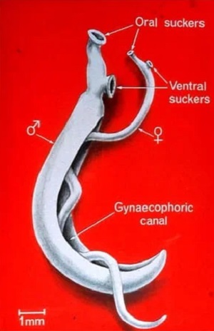
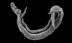
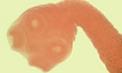
Image: Schistosoma mansoni adult male and female (Source: slidesharecdn)
Adult
the body is covered with integument which protects the fluke from the immune system of the host
unisexual (diecious) – male holds female in the gynecophoral canal
the male is short and stout (1cm-1.5cm in length and 0.9mm in breadth)
female is 2cm in length and 0.25mm in breadth
the female can lay around 100-300 terminal-spined eggs per day
the life span of an adult is 20 years while the parasite lives in man for 5-6 years
the body surface of the male is finely tuberculate
in males, the number of testes varies from 6 to 9 and is arranged in a cluster
the ovary is positioned in the anterior to middle of the female body
about 1-4 eggs are present in the uterus
does not multiply in man
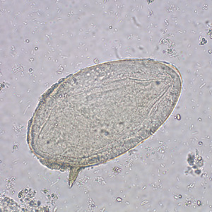
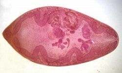
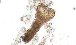
Image: Schistosoma mansoni egg (Source: CDC)
Egg
measures 114μm to 175μm in length and 40μm to 70μm in breadth
elongated, oval-shaped, yellowish brown and non-operculated
identifying feature of Schistosoma mansoni egg is a lateral spine near the rounded posterior end
the sharp lateral spine measures 20μm in length
eggs are fully embryonated when laid
Schistosoma mansoni eggs are short-lived and infectious to snails only
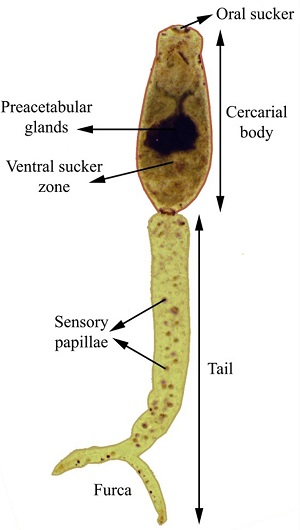
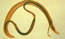
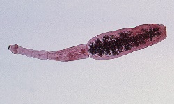
Image: Schistosoma mansoni cercaria morphology (Source: ResearchGate)
Infective form
Cercaria
the infective form of Schistosoma mansoni
elongated oval body with fork-tail
measures 185μm to 230 μm in length and 75 μm to 110 μm in breadth
has two suckers and a bifurcated elongated tail measuring 100 μm in length
the entire body is covered with extremely spine-like projections
short-lived- lives only 24 hours to 72 hours
male and female cercaria are morphologically similar to other Schistosoma species
basophilic in nature with two pairs of cephalic glands