Strongyloides stercoralis - Laboratory Diagnosis
Laboratory diagnosis of Strongyloides stercoralis
The laboratory diagnosis of Strongyloides stercoralis is done by demonstration of larvae in samples such as stool and in disseminated cases in the urine, and sputum.
Sample
stool
urine
sputum
* urine and sputum are collected in cases of disseminated infection
Stool microscopy
only larvae can be observed in the stool and not the adult form during stool microscopy
since the number of eggs released is low i.e. less than 50 eggs per day by a female Strongyloides stercoralis, a single stool examination is not adequate
although has a low sensitivity (30% to 40%), examination of multiple samples (minimum of three) the sensitivity is increased
stool examination is negative in cases of early infection
Strongyloides stercoralis larvae must not be confused with other parasite larvae such as the resembling hookworm larvae
distinguished by a shorter buccal cavity, notched tail, and esophagus which is present in one-half part of the body
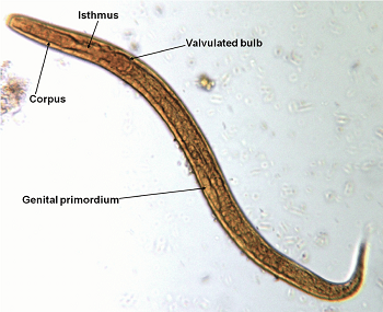
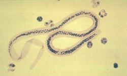
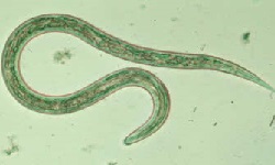
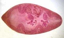
Image: adult parasitic female Strongyloides stercoralis (Source: Creative Diagnostics)
Stool culture
stool culture for diagnosis of Strongyloides stercoralis is done mostly in suspected cases where stool microscopy has given negative results
the concentration of samples can be done by the formalin-ether sedimentation method
stool culture is done by:
* Harada-Mori filter paper method
* Baermann funnel method using charcoal
* agar plate method (done in Petri dishes containing water or solid agar)
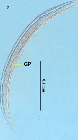
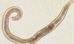
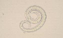
Image: rhabditiform larvae of Strongyloides stercoralis (Source: ResearchGate)
Entero-test
The procedure for the entero-test, a diagnosis for Strongyloides stercoralis, is as follows
also known as string test
a gelatin capsule is tied to the end of a nylon string with a weight attached to it
the patient swallows the capsule while the other free end is taped to the cheek
the capsule is dissolved in the patient’s stomach, releasing the nylon string
since the string is attached to a weight, it travels down to the duodenum and jejunum
after 3-4 hours or overnight, while the patient maintains fasting, the string is pulled out
the bile stained mucus is then collected on a glass slide and immediately observed under a microscope for Strongyloides stercoralis larvae
Serodiagnosis
Numerous serological tests based on antibody detection are used for the diagnosis of Strongyloides stercoralis infection.
The major disadvantage of the serological test is it cannot differentiate between recent and old infections as well as cross-reactions with other parasitic nematodes.
Commonly used serological tests include:
indirect haemagglutination assay (IHA)
Immunofluorescence assay (IFA)
enzyme-linked immunosorbent assay (ELISA) - (sensitivity – 80-90%)
Intradermal skin test
The intradermal skin test is based on the host hypersensitivity reaction against Strongyloides stercoralis antigen. It uses larval extracts as the antigen and was first described by Fulleborn in 1926.
The test shows negative results if the patient undergoes chemotherapy.
The steps for this procedure include:
a required volume of Strongyloides stercoralis antigen is injected intradermally
the injection site is checked for erythematous reaction (positive case)
however, this method is rarely used now due to low sensitivity and specificity
Molecular diagnosis
PCR is one of the most common molecular diagnoses used for the diagnosis of Strongyloides stercoralis.
Imaging methods
Some imaging methods used are:
X-ray
CT-scan