Strongyloides stercoralis - Life Cycle, Pathogenesis, Pathology
Life Cycle of Strongyloides stercoralis
Although the life cycle of Strongyloides stercoralis is completed in a single host, it is complex. The life cycle of the parasite is unique due to its ability to auto-infection and multiply within the host.
The parasite has two distinct life cycles – one within the human body (including autoinfection) and another free-living in the soil.
Principle host: Man is the principal host while dogs, cats, and other animals can serve as reservoirs.
Host cycle
Humans acquire the infection after the infectious form of the Strongyloides stercoralis parasite i.e. the filariform larvae penetrates the skin or buccal mucosa
upon reaching the small blood vessels, these larvae are carried to the heart and then to the lungs via blood circulation
the parasite leaves pulmonary capillaries to reach the alveoli and move upward to the trachea from where they are swallowed back into the intestine – the duodenum and the jejunum
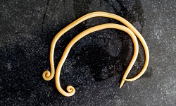
in the intestine, these larvae moult twice to mature into adult female worms
these female Strongyloides stercoralis burrow deep into the intestinal mucosa where they begin lay eggs via parthenogenesis within 28 days
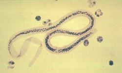
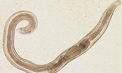
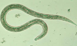
these eggs immediately hatch to release non-infective Strongyloides stercoralis rhabditiform larvae that form
these newly hatched larvae are passed out along with the host stool to follow the free-living cycle
another path of the life cycle is the rhabditiform larvae migrating back to the intestinal lumen where they transform to filariform larvae to cause autoinfection (parasitic cycle)
the autoinfection by Strongyloides stercoralis results due to the non-infective larvae, rhabditiform larvae transforming into the infective filariform larvae while still in the host intestine
similar to other Strongyloides stercoralis infections, in autoinfection, the larvae penetrate the intestinal mucosa resulting in internal autoinfection of the skin of the perineal area resulting in external autoinfection
in the above ways, the cycle is continued in a single host with a repeated migratory cycle
Free-living cycle
If the non-infective rhabditiform larvae present in the faeces reach the soil, the indirect life cycle (new free-living adults) of Strongyloides stercoralis begins or a direct cycle (infective filariform larvae) can take place.
Direct cycle (host-soil-host)
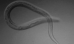
in the soil, the Strongyloides stercoralis rhabditiform larvae undergo two moults to turn into the filariform larvae
the transformation takes place within 3 days to 4 days
these filariform larvae which come to human contact, penetrate the human skin to cause infection and the life cycle is repeated
sexual phase is absent
each rhabditiform larvae gives rise to one single filariform larvae
Indirect cycle
within 24 to 30 hours of hatching of Strongyloides stercoralis rhabditiform larvae, it matures into free-living males and females
the males fertilize the female and the gravid females begin to lay eggs
eggs hatch to release the
within 3 to 4 days, the larvae moult to form the infective Strongyloides stercoralis filariform larvae – each rhabditiform larvae hatching from the first batch of eggs gives rise to 30 filariform larvae
these filariform larvae when coming into human contact, penetrate the human skin to cause infection and the life cycle is repeated
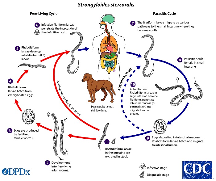
Figure: Strongyloides stercoralis life-cycle (Source: CDC)
Pathogeneis, Pathology of Strongyloides stercoralis
Both the larval and the adult worms of Strongyloides stercoralis are pathogenic to their human hosts.
Pathogenicity of larva
at the site of penetration by filariform larvae, macules and papules are produced
in a sensitized host, the parasitic antigens can result in allergic reactions such as urticaria and pruritus
in the lungs, the migrating larvae produce tissue damage, alveoli injury, and bronchial epithelium injury resulting in bronchopneumonia or pneumonitis
pathological changes include inflammatory exudates of macrophages, epithelial cells, and minimal haemorrhage
Pathogenicity of adult worm
in the host intestinal mucosa (duodenum and jejunum), mild to moderate levels of pathological changes take place
common changes include oedema, cellular infiltration, partial villous atrophy, and in severe cases, ulcers and long-standing fibrosis is seen
in cases of autoinfection and hyper infection syndrome, mucosal inflammation is severe with thickened and oedematous sigmoid colon and rectum