Taenia solium - Introduction, Classification, History, Habitat, Morphology
Introduction of Taenia solium
Taenia solium, also known as pork tapeworm, causes intestinal taeniasis. Infection is acquired orally in humans after ingesting raw or undercooked pork containing the larvae (Cysticercus cellulosae) of the parasite. Pigs acquire the infection after consuming human feces contaminated with the tapeworm egg.
Pig is the usual intermediate/secondary host where larval development takes place while the definitive host of T. solium is the human. However, man can also act as an intermediate host making Taenia solium the only cestode with a man acting as both an intermediate/secondary and definitive host.
The Cysticercus cellulosae causes a serious disease called cysticercosis (especially neurocysticercosis), which is absent in T. saginata.
Classification of Taenia solium
Kingdom: Animalia
Phylum: Platyhelminthes
Class: Cestoda
Order: Cyclophyllidea
Family: Taeniidae
Genus: Taenia
Species: T. solium
History of Taenia solium
In 1854, van Beneden first described the life cycle of Taenia solium. He was able to demonstrate the larvae (Cysticercus cellulosae) in the pig muscles after feeding it eggs from human feces.
A year later, in 1855, Kuchenmeister displayed an adult tapeworm in the intestine of humans.
Habitat of Taenia solium
In the definitive host, the adult tapeworm can be found in the small intestine (upper jejunum). The parasite remains attached to the intestinal mucosa with the help of hooks or rostellum.
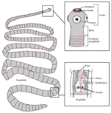
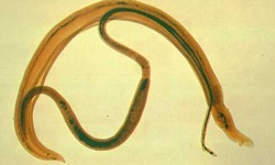
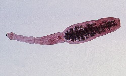
Fig: Taenia solium morphology (Source: MSD Manuals)
Morphology of Taenia solium
Adult worm
white, ribbon-like, flattened, and segmented worm
contains head (scolex), neck, and body (strobila) which consists of a chain of segments (proglottids)
measures 2 meters to 3 meters in length
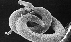
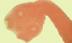
Scolex
round in shape
measures 1mm in diameter
has four suckers
conspicuous hooks or rostellum is present – hence called armed tapeworm
rostellum consists of two rows of alternating small and large hooks
Neck
short and fragile
measures 5 mm to 10 mm in length
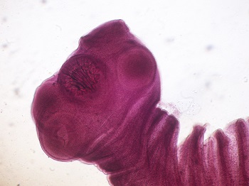
Fig: Taenia solium scolex and neck (Source: Wikipedia)
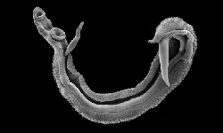
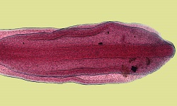
Strobila
measures 2 meters to 3 meters
consists of 800-1000 proglottids or segments
the proglottids are arranged in the linear sequence of immature, sexually mature, and gravid
immature proglottids are broader than longer
mature proglottids are also wider than long, similar to that of T. saginata
gravid proglottids are longer than broad (measure 12mm x 6mm), contain around 30,000 to 50,000 eggs in each segment
gravid proglottids are grayish-black in color and turn transparent when fully developed
common genital opening is present laterally in the middle of each segment with alternation between the left and right side
after breaking off from Strobila, around 5-6 gravid segments are passed passively in chains along with the feces
Taenia solium can live for 25 years and the most single worm is found infecting a human- multiple infections are rare
The proglottids of Taenia solium are different from that of T. saginata as Taenia solium proglottids:
presence of 150-200 testes
the ovary has a third accessory lobe
the ovary is present on the posterior side of the segment
vaginal opening lacks a muscular sphincter
has a median longitudinal uterine stem with 7-13 branches on each side
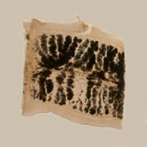
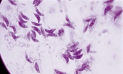
Source: Taenia solium - unstained gravid proglottids (Source: cych.org)
Egg
round or oval in shape
measures 41μm to 43μm in diameter
infective to pigs as well as humans
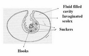
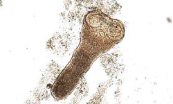
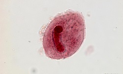
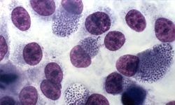
Fig: Taenia solium Cysticercus cellulosae (Source: pathologyoutlines)
Infective Form
Cysticercus cellulosae
also known as taenia cyst of Taenia solium
the infective form of the parasites for humans
this larval form is found in the muscles of both pigs and man
small, oval in shape measuring 3mm x 15mm
are fluid-filled milky white bladder-like structures rich in albumin and salts, with a translucent wall through which a single dense white body (invaginated scolex) can be seen
cross-section of the scolex shows several layers of folded smooth muscles with parts of suckers or hooklets
cyst wall measures 100μm -200μm and are composed of three layers
* inner layer consists of longitudinal and circular muscles
* middle layer consists of pseudoepithelial cells
* outer cuticular later contains a dentate membrane with a microvillus projection responsible for interaction with host tissues
cyst and host tissue are separated by a thin collagenous capsule