Trichinella spiralis - Laboratory Diagnosis, Treatment, Prevention, Control
Laboratory diagnosis of Trichinella spiralis
The laboratory diagnosis of Trichinella spiralis is done by demonstration of first-stage larvae, or adults worm (rare occasion) of the parasite.
Samples
biopsy (muscles)
* larvae is found in muscles after 3rd of 4th week of infection
Microscopy
The microscopy of Trichinella spiralis includes:
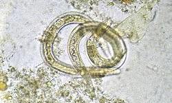
Demonstration of larva
direct slide technique used to demonstrate the Trichinella spiralis larva
* in this procedure, sample muscle tissue is pressed between two glass slides
* examined under a microscope for presence/absence of larvae under low power microscope
viable larvae can be demonstrated by dissolving the muscle sample with a 1% pepsin-hydrochloric acid mixture and examining under 10x objective
presence of viable larvae indicates recent infection
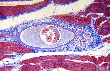
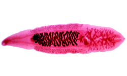
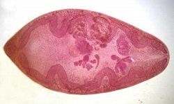
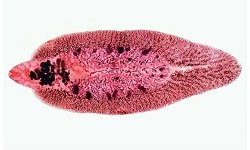
Image: Trichinella spiralis larvae inside muscle tissue (Source: Encyclopedia Britannica)
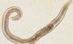
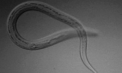
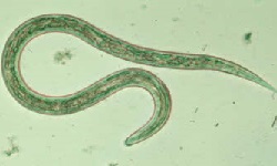
Demonstration of adult worm
adult Trichinella spiralis are rarely seen in the feces
if demonstrated in the feces, they are mostly degenerated and not recognizable
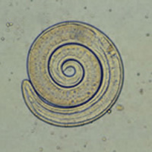
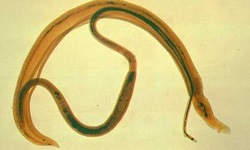
Image: adult Trichinella spiralis (Source: CDC)
Serodiagnosis
Numerous serological tests based on antibody detection are used for the diagnosis of Trichinella spiralis infection. These tests involve the use of homogenates retrieved from muscle larvae or excretory-secretory (ES) as antigens. Since the TSL-1 group of larcal secretory antigens is present in all species of Trichinella, it can be used for diagnosis.
Commonly used serological tests include:
bentonite flocculation (sensitivity – 90%)
latex agglutination
fluorescent antibody test
parasite-specific indirect immunoglobulin G (IgG) enzyme-linked immunosorbent assay (ELISA)
Antibody level peaks in the second or third month after infection which slowly decline for several years. During the acute stage of trichinellosis, antibody levels remain undetectable until 3 to 5 weeks after infection.
Intradermal skin test
The intradermal skin test, which is based on the host hypersensitivity reaction, is developed by Bachman. This test shows positive results 11 to 16 days after infection.
The steps for this procedure include:
0.1 ml of 1 in 10,000 dilutions of Trichinella spiralis antigen, also known as Bachman antigen, is injected intradermally
the injection site is checked for immediate erythematous reaction (positive case) within 15 minutes to 20 minutes
however, this method is rarely used now due to low sensitivity and specificity
Molecular diagnosis
PCR is one of the most common molecular diagnoses used for the diagnosis of Trichinella spiralis.
Imaging methods
X-ray
CR-scan
Treatment of Trichinella spiralis
Antihelmenthic drugs such as thiabendazole and mebendazole can be used for the treatment of Trichinella spiralis. Corticosteroid treatment is also done to reduce the immunologic response.
Prevention, and Control of Trichinella spiralis
The prevention and control of Trichinella spiralis can be achieved by:
deep-freezing meat products at -15° C for 20 days or at -30° C for 6 days
cooking at 70° C or above kills the larvae in meat products
avoid consumption of smoking, curing, or drying of meat
not eating raw or undercooked pork
avoid feeding garbage to pigs