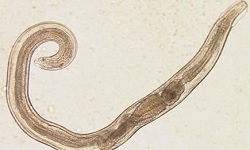
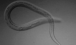
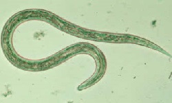
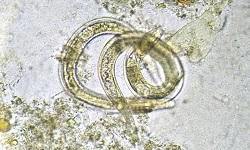
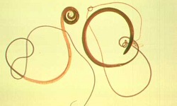
Trichinella spiralis - Life Cycle, Pathogenesis, Pathology
Life Cycle of Trichinella spiralis
The life cycle of Trichinella spiralis occurs in a single host which serves both as the definitive host and intermediate host i.e. a single host can harbor the adult as well as larva. However, to complete the life cycle, two different hosts are required.
Primary host: Pig
Natural hosts: carnivores, omnivorous animals including rodents, bears, and hyenas. Humans, which is an accidental hosts, are the dead end for Trichinella spiralis.
Humans acquire the parasitic infection after consumption of raw or insufficiently cooked pork harboring viable encysted larvae
the cyst wall is digested in the host stomach, freeing the Trichinella spiralis larvae
the larvae penetrates the duodenal and jejunal mucosa
further development of the larvae into males and females takes place within 2 days
mating and fertilization take place within the intestinal mucosa as an intracellular parasite
females burrow deep into the intestinal wall or mesenteric lymph nodes
Trichinella spiralis females burrow deep into the host intestinal wall and within 6-7 days, deposit the larvae
deposition of the larvae continues for several weeks
each female can lay 1,500 to 2,000 newborn larvae during its lifespan
male Trichinella spiralis die shortly after mating while the females die shortly after completion of laying larvae
the newborn Trichinella spiralis larvae penetrate the mesenteric and lymphatic venules
the larvae are carried by general circulation to various host organs and tissues – with a predilection for muscles of the tongue, diaphragm, eye muscle, masticatory muscles, muscles of arms and legs, and masticatory muscles
inside the host muscle tissues, the larvae grow with an increase in size and encyst within 3 weeks
in dead-end hosts like humans, calcification of the dead larvae begins in 6 to 18 months
in other sites such as the myocardium, and central nervous system, dead larvae do not encyst
transmission occurs after ingestion of raw or undercooked meat infested with viable cysts or larvae
although infection in man is a dead end, the life cycle of Trichinella spiralis is maintained in nature
primary host (pig) acquires infection after ingestion of other pig carcasses (pig-to-pig), rats (rat-to-pig)
rats also acquire infection from infected rats (rat-to-rat) or from pigs (pig-to-rat)
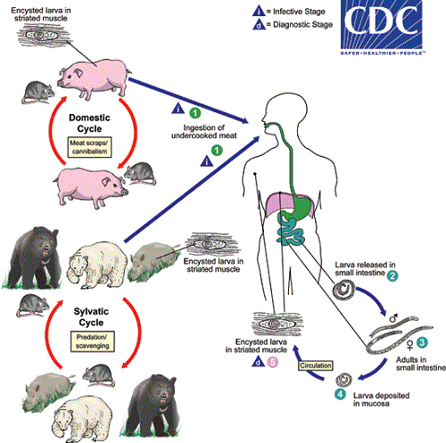
Figure: Life cycle of Trichinella spiralis (Source: CDC)
Pathogeneis, Pathology of Trichinella spiralis
Both adult worms and larvae are pathogenic. Adult female Trichinella spiralis inhabitant the intestine results in gastrointestinal syndromes while migrating larvae cause allergic manifestations such as fever, eosinophilia, and edema of the face. Moreover, the encysted larvae present in the skeletal muscles cause muscular pain
Pathogenicity of adult worm
adult Trichinella spiralis females present in the small intestine results in acute eosinophilia, inflammatory reactions, hyperemia, edema
Pathogenicity of migrating larva
migrating Trichinella spiralis larvae cause profound lesions in the skeletal muscles such as myositis, basophilic granular degradation of muscle fibers
associated with hyperemia, edema, hemorrhage
in the muscle fiber cells, the larvae can induce a number of changes including:
* hypertrophied nuclei
* inactivation of transcription of muscle genes
* reduced expression of certain muscle-specific proteins
* infected muscle cell ceases to develop as it becomes arrested in the reproduction cycle of the cell at the gap 2 or M phase
Pathogenicity of encysted larva
encysted larvae occur only in skeletal muscles
since encapsulation of the Trichinella spiralis takes place between 21 to 90 days, pathological changes including the formation of a lemon-shaped sheath around the larva
other clinical manifestations include fever, myalgias
within 6 months to 2 years, the intramuscular cysts calcify
although the growth or encapsulations do not take place in the myocardium, the encysted larva of Trichinella spiralis produces inflammation, necrosis, and fibrosis of myocardial fibers
in the central nervous system, these larvae produce granulomatous nodules, and vascularisation of small arteries as well as capillaries which leads to frequent brain hemorrhage