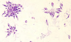
Trypanosoma brucei complex - Epidemiology, Transmission, Lab Diagnosis, Treatment, Control
Epidemiology of Trypanosoma brucei complex
Epidemiologically, the Trypanosoma brucei complex is prevalent in 36 African countries of sub-Sahara where tsetse fly is present.
West African trypanosomiasis is endemic to Central and Western African countries such as Congo, Angola, Northwest Sudan, Nigeria, Guinea, Uganda, etc.
East African trypanosomiasis is endemic to Southern and Eastern African countries including Uganda, Kenya, Malawi, Zambia, Tanzania, and Mozambique.
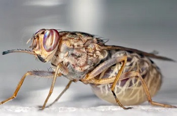
Image: Trypanosoma brucei vector - tsetse fly (Source: Entomology Today)
Reservoir, Source, Transmission of Trypanosoma brucei complex
Since East African trypanosomiasis is a zoonotic disease, the sources and reservoirs of Trypanosoma brucei rhodesiense include wild animals (antelope- bushbuck, hartebeest), and domestic animals such as cattle. Vectors for transmission of his disease are G. pallidipes, and G. morsitans, and is seen mostly in hunters, honey, and firewood collectors.
West African trypanosomiasis is not a zoonotic disease. The source and reservoir of Trypanosoma brucei gambiense is an infected man. Transmission takes place by the tsetse fly (G. palpalis, G. tachinoides, G. fuscipes) and infection is primarily seen in rural areas.
Laboratory diagnosis of Trypanosoma brucei complex
The laboratory diagnosis of Trypanosoma brucei complex begins with the collection of samples:
Sample
blood
cerebrospinal fluid
chancre fluid
lymph node aspirate
bone marrow
Microscopy
Wet mount method
The wet mount method microscopy is mostly done for chancre fluid and lymph node aspirate
demonstration of highly motile trypanosomes confirms the Trypanosoma brucei infection
a hundred microscopic fields at 40x must be done before concluding the sample is negative
samples are also stained by Giemsa stain and fixed to examine the parasite under the microscope
Blood smears
both thick and thin blood smear microscopy stained by Giemsa stain is done
less sensitive procedure as the parasites are easily disrupted during the preparation of the smear
Wright and Leishman's stains are not used
Concentrated blood microscopy
Concentrated blood microscopy is done by hematocrit centrifuge technique or DEAE cellulose anion exchange chromatography
useful in the detection of trypanosomes in the blood specimen
* if the sample is CSF, double centrifugation followed by is a simple and sensitive method
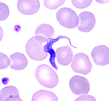
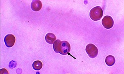
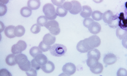
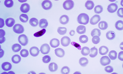
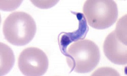
Image: Trypanosoma brucei slender trypomastigote microscopy (Source: Wikipedia)
Culture
The culture of the Trypanosoma brucei complex is done by animal inoculation or via in-vitro culture.
Animal inoculation
if the microscopy of blood, lymph fluid, and CSF samples fail to detect the trypanosomal parasites, inoculation in laboratory mice and rats is done
several mice are inoculated with the sample intraperitoneally and from the 3rd day to the 21 days, tail vein blood is collected and examined
not useful in the detection of Trypanosoma brucei gambiense
In-Vitro culture
Trypanosoma brucei gambiense and Trypanosoma brucei rhodesiense can be cultured in-vitro in the NNN media and the Tabies’ biphasic media
can also be grown in primary murine bone marrow culture (feeder layer) in RPMI 1640, Eagle’s minimum essential medium (MEM) which has been supplemented with 20% fetal calf serum (FCS) and salts
in such cultures, the monomorphic forms of the parasite can be isolated inside the feeder layer while pleomorphic forms are found in the liquid medium
However, as the process is slow, this is not done in routine diagnosis
Serodiagnosis
commonly used serological methods include indirect haemagglutination (IHA), Indirect immunofluorescent antibody (IFA), Direct Agglutination test (DAT), Enzyme-Linked Immunosorbent Assay (ELISA)
specific antibodies are detected within 2 weeks to 3 weeks from infection
CATT is done for field use and mass screening for Trypanosoma brucei complex with 96% sensitivity
Molecular methods
The molecular methods used for diagnosis of Trypanosoma brucei complex includes:
PCR
DNA probes
Imaging methods
In the later stages of the disease caused by Trypanosoma brucei complex,
CT scan of the brain shows cerebral edema
MRI shows white matter enhancement
Treatment of Trypanosoma brucei complex
If the disease is in the early stage, pentamidine and arsenical suramin are the drugs of choice for treatment
no drugs are available to prevent the transmission of parasites via blood transfusion
Prevention, Control of Trypanosoma brucei complex
The prevention, control of Trypanosoma brucei complex include as follows:
reduction of the tsetse fly population by using insecticides
evade direct contact with potential animal reservoirs
use of bed nets, window nets, or insect repellents
Screening of blood donors