Brugia malayi - Culture, Life Cycle
Culture of Brugia malayi
The culture of Brugia malayi can be achieved via media culture or via animal culture.
Culture media
In-vitro tissue culture of L3 larvae to L4 larvae and juvenile adult Brugia malayi can be done using rhesus monkey kidney cell lines (LLC-MK2) which has been supplemented with RPMI-1640 medium and 10% inactivated human serum.
The inoculated media is then incubated at 37°C in the presence of air.
Laboratory animals
Brugia malayi is the only pathogenic filarial parasite that can cause infection in small laboratory animals such as rodents including congenital athymic and immuno-deficient nude mice. Other animals (mongolian jirds, ferrets, dogs, cats) are tested to study the pathogenesis of lymphatic filariasis.
In these animals, microfilariae are present in the peripheral blood for a longer duration.
For the culture of Brugia malayi in laboratory animals, 200-300 L3 larvae are spontaneously inoculated on the thigh. After a pre-patent period of 6 weeks, microfilariae are demonstrated in the peripheral blood.
Life Cycle of Brugia malayi
The life cycle of Brugia malayi is completed in two classes of hosts – the definitive host and the intermediate host.
Definitive host:
Nocturnal periodic Brugia malayi – Humans
Sub-periodic Brugia malayi – humans, leaf monkeys (Presbytis spp.), macaques (Macacca spp.), cats
Intermediate host: Mosquitoes
* nocturnal periodic Brugia malayi transmitted by Anopheles and Mansonia mosquitoes
* sub periodic Brugia malayi transmitted by Mansonia and Coquilletcidia
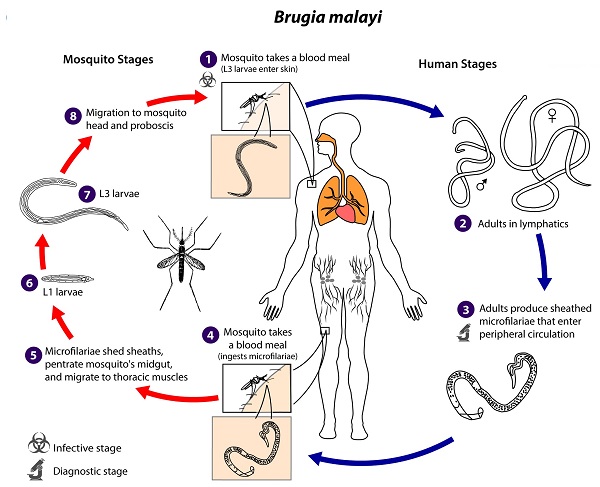
Brugia malayi life-cycle (Source: CDC)
Transmission of Brugia malayi begins with the bite of infected mosquitoes onto a healthy person.
bite of an infected mosquito transmits the third-stage larvae (L3) onto the skin
the larvae enter through the punctured wound made by the infected vector and reach the peripheral lymphatic vessels
Brugia malayi larvae quickly migrate to inguinal lymph nodes where they metamorphose and grow into sexually mature adult males and adult female
both adult male and female filarial nematodes live in regional lymphatic vessels and nodes (mostly in the cortex of lymph nodes and testicular tissues) as coiled-up structures
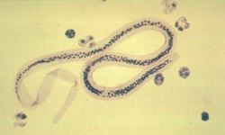
after fertilization, the ovo-viviparous female deposits microfilariae
the newly emerged microfilariae appear in the peripheral blood within 8 to 12 months of infection
these microfilariae circulate in the blood from 6 months to 2 years
if these microfilariae are not taken up by the vector (mosquitoes), they die
if the microfilariae are ingested by the vector, within 2 to 6 hours inside the vector’s stomach, they lose their sheath
within 4 hours to 17 hours, these microfilariae larvae migrate to the thoracic muscles
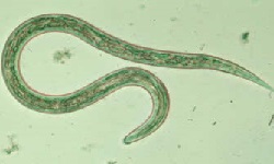
the slender Brugia malayi microfilariae larvae undergo development in the next 48 hours to molt into first-stage larvae (L1)
these L1 larvae are thick, short, sausage-shaped with spiky tails, measuring 124 to 250 µm in length and 10 to 17 µm in breadth
Between 3rd day to 7th day, the L1 larvae of Brugia malayi molt to become larger sausage-shaped second-stage larvae (L2)
L2 larvae measure 225 to 300 µm in length and 15 to 30 µm in breadth
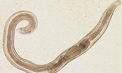
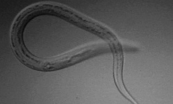
the L2 larvae eventually moults to third-stage larvae (L3) by the 10th and 11th day
on the 14th day of infection, L3, the infective form of the parasite, migrates to the salivary glands of the mosquito vector
if the vector takes a blood meal from a healthy person, the L3 Brugia malayi larvae are released from the tip of the proboscis of the infected mosquito into a new host
*development of Brugia malayi larvae in mosquitoes is completed in 10 to 20 days.
* microfilariae of the filarial nematode do not undergo multiplication to increase in number i.e. only each microfilaria develops into one infective larva
.jpg)